
Microbiology is the scientific study of microorganisms and related microscopic infectious agents: their structures, growth, genetics, environments, interactions with hosts and effects on health. For nursing students, microbiology explains why hand hygiene works, how infections may spread, how laboratory tests contribute to clinical reasoning, and why specimen handling matters. It is not exclusively a study of disease: microorganisms also contribute to digestion, food fermentation, environmental nutrient cycles and biotechnology.
This comprehensive introductory chapter covers definitions, branches, microbial groups, the history of microbiology, the experimental foundations of germ theory, and practical implications for infection prevention. It provides educational principles, not independent authority to diagnose infections or perform clinical procedures.
Learning objectives
- Define microbiology and distinguish microorganisms from pathogens and infectious agents.
- Compare bacteria, archaea, fungi, protozoa, viruses, helminths and prions.
- Name the major branches of microbiology and describe how each contributes to healthcare.
- Describe why microscopy transformed science and identify key work by Leeuwenhoek, Pasteur and Koch.
- Explain how controlled experiments challenged spontaneous generation.
- Summarise germ theory, Koch’s postulates and the limitations of applying them universally.
- Apply introductory microbiology to infection prevention, specimen quality and safe nursing communication.
1. Definitions: microbiology, microbes and pathogens
A microorganism is usually defined as a biological organism whose individual form requires magnification for detailed observation. Most bacteria are microscopic single-celled organisms. Many yeasts and protozoa are also unicellular, while certain fungi can form multicellular structures. Viruses are acellular: they consist of genetic material and other components but are not cells, and they require host-cell machinery for replication. Adult parasitic worms may be visible to the naked eye, but their microscopic eggs and larvae make them part of medical parasitology.
The terms microbe, pathogen, normal microbiota, colonisation and infection are not interchangeable. A pathogen is an organism or agent capable of causing disease in suitable circumstances. Microbiota are microbial communities associated with a host site, such as the skin or gastrointestinal tract. Colonisation means that microbes are present and may reproduce at a site without necessarily causing illness. Infection involves a host–agent interaction that may result in tissue damage or symptoms; asymptomatic infection also occurs.
Microbial pathogenicity is contextual. An organism that is common in the intestine might cause trouble if it reaches a normally protected body site. Exposure does not always lead to colonisation, infection or disease. Host immunity, the entry route, the number of organisms involved and their biological properties all influence what happens.
2. Major types of microorganisms and infectious agents
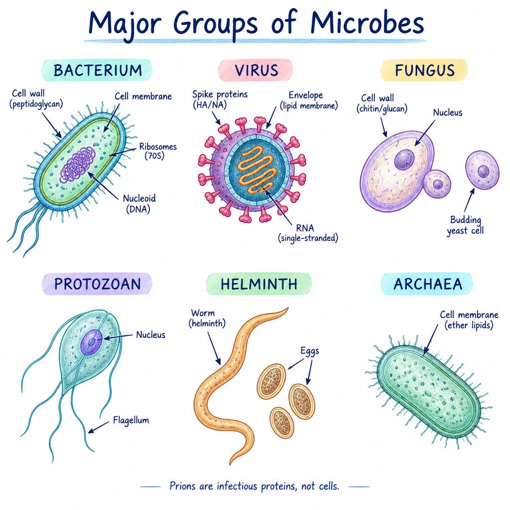
| Group | Structure and reproduction | Example and nursing relevance |
|---|---|---|
| Bacteria | Prokaryotic cells; many divide by binary fission. | Escherichia coli includes harmless gut-associated strains and strains associated with illness. |
| Archaea | Prokaryotes with molecular and membrane features distinct from bacteria. | Present in diverse environments, including human-associated communities. |
| Fungi | Eukaryotic yeasts or multicellular moulds. | Candida species may colonise mucosal surfaces or cause infection in particular circumstances. |
| Protozoa | Mostly unicellular eukaryotes. | Giardia is associated with intestinal infection. |
| Viruses | Acellular particles requiring living host cells to replicate. | Influenza virus illustrates the relevance of respiratory infection prevention. |
| Helminths | Multicellular parasitic worms; some life stages are microscopic. | Parasitic roundworms and tapeworms are studied in medical parasitology. |
| Prions | Infectious misfolded proteins without nucleic acid. | Associated with rare transmissible spongiform encephalopathies. |
Not all these categories are living cellular organisms. In particular, viruses and prions should not be described as ordinary cells. Different microbial groups require different laboratory approaches and, where treatment is indicated, different therapeutic strategies.
3. Main branches of microbiology
| Branch | What it studies | Nursing application |
|---|---|---|
| Bacteriology | Bacteria and their biology | Understanding bacterial culture and antibiotic-susceptibility reports |
| Virology | Viruses and replication | Viral infection prevention and health education |
| Mycology | Fungi and fungal disease | Understanding risk in people with impaired immune defences |
| Parasitology | Protozoan and helminth parasites | Understanding water-, food- and vector-associated illness |
| Immunology | Host immune responses | Vaccines, susceptibility and inflammatory responses |
| Clinical microbiology | Detection and interpretation of microbes in clinical specimens | Specimen identification, correct collection and communication |
| Microbial genetics | Inheritance and transfer of microbial characteristics | Understanding antimicrobial resistance |
| Public-health microbiology | Transmission and microbial patterns across populations | Surveillance, outbreak response and prevention |
| Environmental and food microbiology | Microbes in ecosystems, water and food | Sanitation, food safety and community education |
These branches are interdependent. A respiratory outbreak investigation may involve virology, specimen testing, epidemiology, immunology and infection-prevention practice simultaneously. The purpose of this classification is to clarify the questions a specialist asks—not to suggest that healthcare disciplines work in isolation.
4. The period before microscopes
People encountered microbial effects long before recognising microbes themselves. Bread rising, food fermentation, spoilage and epidemic disease were visible phenomena, but the causes were largely invisible. Historical explanations included humoral theories, environmental influences and the miasma hypothesis. Although many explanations proved incomplete, observations about water, crowding and hygiene helped inspire later scientific enquiry.
Robert Hooke (1665) described structures observed under a compound microscope and introduced the term cell after viewing cork. Microscopes transformed biology by allowing investigators to inspect structures directly rather than inferring their existence only from indirect effects. However, observing cellular structures did not yet prove that particular microorganisms caused particular human diseases.
5. Antonie van Leeuwenhoek: the microscopic world becomes visible
Antonie van Leeuwenhoek (1632–1723) was a Dutch observer who used carefully crafted single-lens microscopes. In the 1670s he described tiny organisms in water and other samples, calling them “animalcules.” His letters and drawings helped establish that a rich world of microscopic living forms existed beyond ordinary human vision.
His contribution was principally observation and documentation. His microscopes permitted direct study of living microscopic forms, but visualising microbes was not equivalent to establishing a causal link to disease. Researchers still needed reliable culture methods, experimental controls, taxonomic frameworks and repeatable disease observations.
Nursing connection: Microscopy can reveal morphological features, arrangements or the presence of organisms. It cannot, without interpretation and appropriate additional testing, prove why a particular patient is unwell. A stained specimen, for example, needs to be understood alongside specimen type, collection quality and the patient’s clinical findings.
6. Louis Pasteur and the end of ordinary spontaneous-generation theory
Spontaneous generation was the historical belief that organisms could routinely develop directly from nonliving material—for example, that microbes simply appeared in broth. Francesco Redi and Lazzaro Spallanzani challenged variations of this belief through experiments, but debate persisted regarding heating, air and contamination.
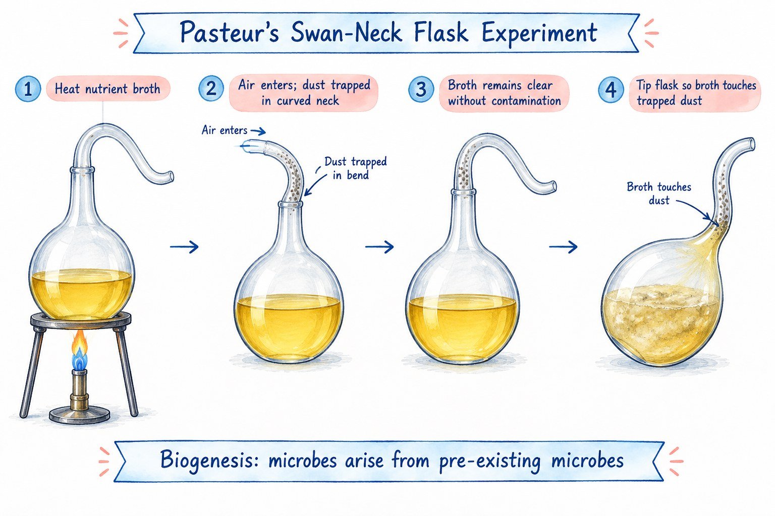
Louis Pasteur (1822–1895) developed evidence through studies of microbial fermentation and the famous swan-neck flask experiments. Nutrient broth was heated under experimental conditions. The flask’s curved neck allowed air to reach the broth but trapped many airborne particles. When contamination was excluded, the broth could remain free of visible growth; when particles carrying microorganisms reached it, growth could occur. This supported the principle that microbes in the broth arose from pre-existing microorganisms rather than appearing spontaneously under those conditions.
- Prepare nutrient broth and heat it as required by the experiment.
- Keep an intact curved flask neck that permits air access while limiting particulate contamination.
- Observe the broth while particles are excluded.
- Allow the broth to contact a source of trapped microorganisms.
- Compare subsequent growth patterns to evaluate contamination as the cause.
Pasteur’s fermentation studies demonstrated that living microorganisms drive particular biochemical processes. His work also contributed to pasteurisation—controlled heating that reduces selected microbial hazards and spoilage—and to the development of vaccines. Pasteurisation is not the same as sterilisation, and product-specific standards determine its practical use.
7. Robert Koch and the scientific evidence for specific causes
Robert Koch (1843–1910) advanced disease microbiology through work on anthrax, tuberculosis and cholera, and by refining laboratory approaches to studying specific microorganisms. The central achievement was not the claim that all disease comes from microbes; it was the development of methods to connect a defined organism with a defined infectious disease.
Koch’s classical postulates are often taught as four related ideas: the suspected microorganism is consistently associated with a particular disease; it can be isolated and studied; it produces comparable illness when introduced into a susceptible experimental host; and it can subsequently be identified again. The framework encouraged investigators to separate plausible associations from stronger evidence of causation.
The postulates are historically influential but not universal rules. Some pathogens cannot be grown through routine methods; viruses require living host cells to replicate. Some people carry organisms without symptoms, host vulnerability varies, and experimentally exposing people to disease would often be unethical. Modern causal evidence also incorporates sequencing, microbial genetics, epidemiology and immunology.
8. Germ theory and the rise of infection prevention
Germ theory recognises that specific microorganisms can cause specific infectious diseases. Its development helped establish modern sanitation, antisepsis, infection prevention, laboratory diagnosis and targeted treatment. It does not say that every organism causes illness, that every disease is infectious, or that every positive test demonstrates active infection.
Ignaz Semmelweis showed the importance of hand disinfection in reducing puerperal fever in nineteenth-century obstetric care. Joseph Lister advanced antiseptic surgical methods. John Snow used epidemiological evidence to investigate cholera transmission in London. These contributions illustrate how observation, patient outcomes, field investigation and experiments complemented laboratory discovery.
9. Historical discoveries at a glance
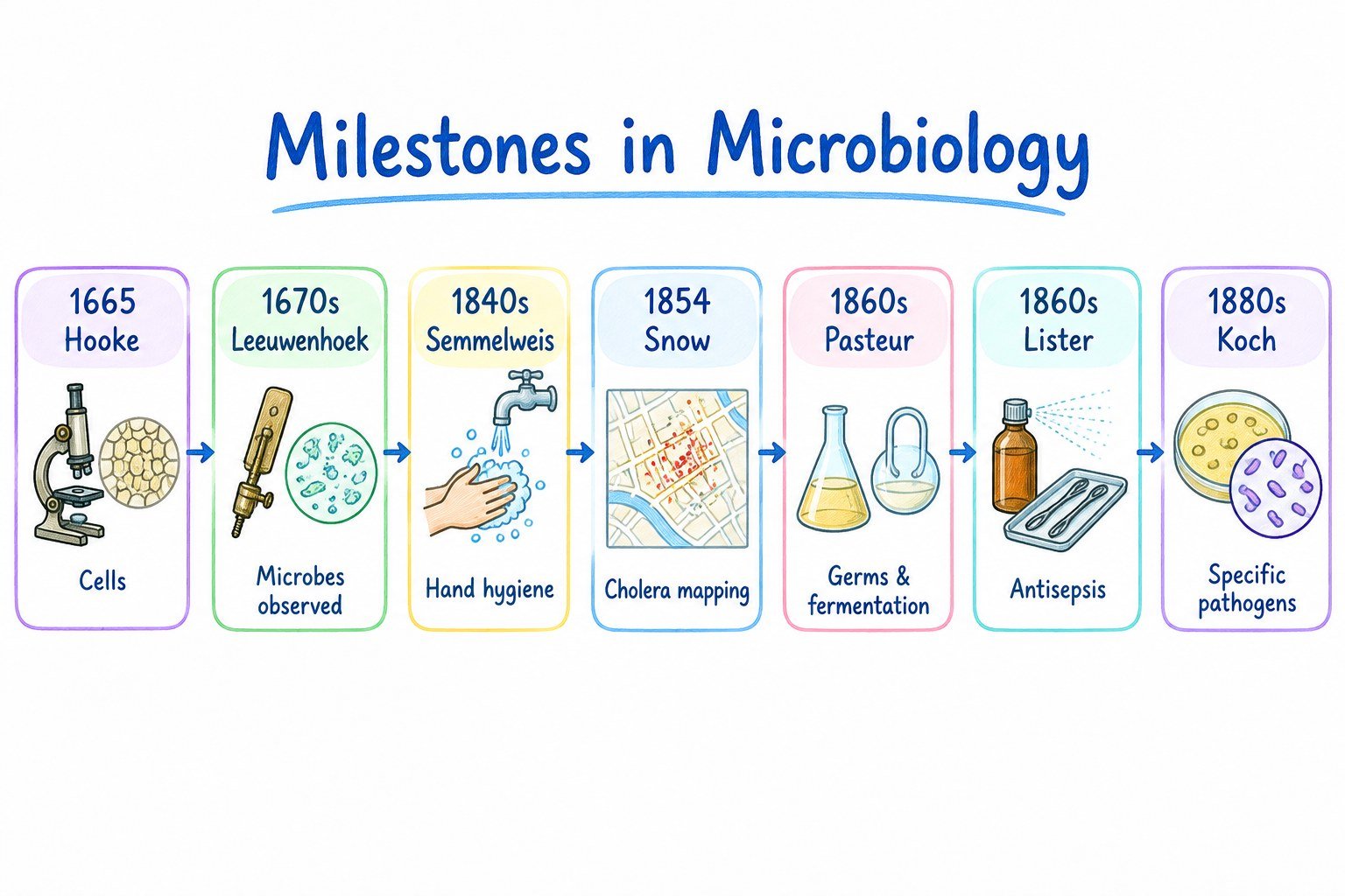
| Period | Scientific advance | Lasting lesson |
|---|---|---|
| 1665 | Robert Hooke’s microscopic study and naming of cells | Microscopy reveals structures invisible to the unaided eye. |
| 1670s | Van Leeuwenhoek’s descriptions of microscopic living forms | Microbes can be directly observed. |
| 1840s | Semmelweis’s hand-disinfection observations | Prevention practices can affect patient outcomes. |
| 1854 | Snow’s London cholera investigation | Population-level evidence helps identify exposure routes. |
| 1850s–1860s | Pasteur’s fermentation and swan-neck flask work | Controlled studies distinguish contamination from spontaneous appearance. |
| 1860s | Lister’s antiseptic surgery | Controlling microbial contamination reduces surgical risk. |
| 1870s–1880s | Koch’s work on specific infectious causes | Causal claims require converging evidence. |
| Modern era | Vaccines, antimicrobial drugs, molecular diagnostics and genomics | Clinical decisions combine microbial, host and public-health evidence. |
10. Why microbiology matters in professional nursing
Infection prevention and patient safety
Healthcare workers interact with patients, body fluids, reusable equipment and shared environments. Understanding microbial reservoirs and transmission explains why hand hygiene, environmental cleaning, respiratory hygiene and appropriate protective equipment are central to preventing avoidable transmission. Standard Precautions apply to all patient care; additional transmission-based measures depend on the situation and local guidance.
Observation without overdiagnosis
Fever, respiratory changes, tenderness or abnormal laboratory values may prompt concern but do not independently identify a causative pathogen. Nurses document objective findings, recognise changes from baseline, communicate clearly and follow escalation procedures. Microbiology informs clinical reasoning without replacing the full assessment performed by the qualified team.
Specimen collection and laboratory interpretation
Correct identifiers, specimen choice, collection technique, timing and transport all matter. A contaminated sample may lead to misleading growth. A positive culture may reflect infection, colonisation or contamination, depending on clinical context. A negative result can also have limitations arising from timing, prior treatment and test sensitivity.
Antimicrobial stewardship
Antibiotics act against susceptible bacteria and are not routinely useful for viral illnesses. Their use depends on clinical diagnosis, local guidelines, likely organisms, susceptibility patterns and patient-specific factors. Nurses support safe administration, monitoring and appropriate education according to professional scope and institutional policy.
Public and community health
Microbiology provides scientific foundations for vaccination, food and water safety, responsible antimicrobial use, respiratory hygiene and outbreak control. Communication should be accurate, respectful and specific to the patient’s needs rather than frightening or blaming people for exposure.
11. The chain of infection explained
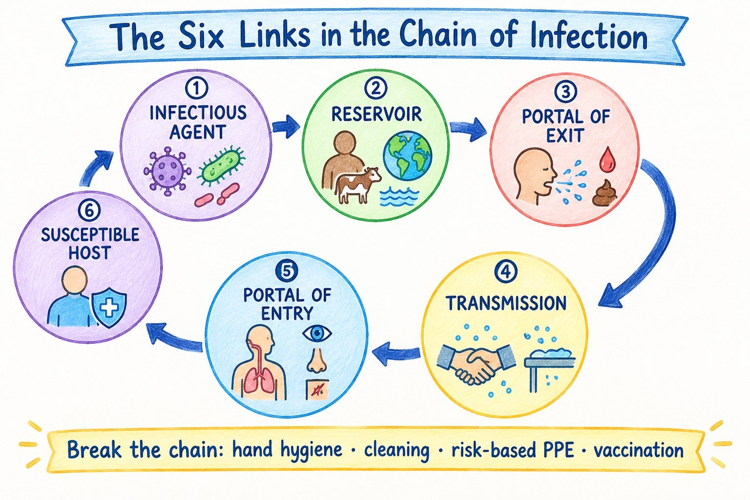
An educational model describes six connected elements: infectious agent → reservoir → portal of exit → mode of transmission → portal of entry → susceptible host. Breaking one or more links can reduce transmission risk. These links are not proof that an exposed person will develop symptoms.
| Link | Meaning | Illustrative control |
|---|---|---|
| Agent | Microorganism capable of causing infection | Validated cleaning and appropriate treatment measures |
| Reservoir | Location in which an organism survives | Managing contaminated surfaces and equipment |
| Portal of exit | Route leaving the source | Respiratory hygiene and safe fluid handling |
| Transmission | How an organism moves to another site or person | Hand hygiene and risk-based PPE |
| Portal of entry | Route into the new host | Aseptic practice and barrier protection |
| Susceptible host | Host factors affecting vulnerability | Vaccination, risk assessment and preventive care |
12. Case-based clinical learning
Case A: gloves are not a substitute for hand hygiene
In a simulated dressing change, a student touches a shared bedside phone and immediately puts on gloves to handle supplies. They argue that gloves make hand hygiene unnecessary. Explain what is wrong. Answer: Gloves can be contaminated during application or use and do not replace indicated hand hygiene. The incident demonstrates a risk that should be corrected, not proof that a patient has become infected.
Case B: positive urine culture but unclear symptoms
A student’s practice notes interpret any bacterial growth in a urine culture as a confirmed urinary tract infection. Answer: Bacteriuria can be asymptomatic, and specimen contamination is possible. The healthcare team must interpret results alongside signs, symptoms and collection quality. The student should document the reported laboratory finding without independently asserting a diagnosis.
13. Fifteen knowledge-check questions and explained answers
- What does microbiology study? Microorganisms and related agents, including beneficial and harmful forms.
- Are viruses cells? No; they are acellular and require host cells for replication.
- Which branch studies fungi? Mycology.
- Which branch studies bacterial organisms? Bacteriology.
- Who first described microscopic “animalcules” during the seventeenth century? Antonie van Leeuwenhoek.
- What did Hooke contribute? Microscopic observations and the term “cell”.
- What did Pasteur’s swan-neck flask experiment challenge? Ordinary spontaneous generation under the conditions tested.
- Why is pasteurisation not the same as sterilisation? It reduces selected microbial hazards but is not designed to eliminate all microbial life.
- Who is associated with classical causal criteria for microbial disease? Robert Koch.
- Why do classical postulates have limitations? Some organisms are not conventionally culturable, and asymptomatic carriage and ethical constraints complicate experiments.
- Does a positive culture always mean active disease? No; colonisation and contamination may explain the result.
- What precautions apply to every patient? Standard Precautions.
- Can gloves replace hand hygiene? No.
- Do antibiotics treat most viral infections? No; antibiotics target susceptible bacteria, not viruses.
- Why are specimen labels critical? They connect laboratory results to the correct patient and specimen.
14. Revision checklist
- Define microbe, pathogen, colonisation, infection and contamination.
- Name at least six microbial or infectious-agent groups.
- Give one healthcare use for five branches of microbiology.
- Compare Leeuwenhoek’s microscopy, Pasteur’s experiments and Koch’s causal investigations.
- Explain the swan-neck flask experiment using an ordered flowchart.
- Identify a limitation of interpreting culture findings without a clinical history.
- Apply the six links in the chain of infection to a simulated nursing scenario.
Next chapter: Chapter 2 — Classification of Microorganisms for Nurses: understand bacteria, archaea, fungi, parasites, viruses and prions and how their differences affect clinical microbiology.
References and further reading
- OpenStax Microbiology — What Our Ancestors Knew.
- OpenStax Microbiology — Types of Microorganisms.
- OpenStax Microbiology — Spontaneous Generation.
- CDC — Core Infection Prevention and Control Practices.
- CDC — Standard Precautions for All Patient Care.
Academic review: This chapter is an original learning resource pending clinical and editorial review. It is not a prescribing, diagnostic or procedural protocol. Always use current institutional guidance and qualified clinical supervision.