
Part 1: Microbiology Foundations
Microorganisms · Bacterial cells · Gram staining · Growth curves · Normal flora
Part 2: Clinical Microbiology
Chain of infection · Precautions · Sterilization · Lab testing · Immunity
Microbiology for nursing students is the study of microorganisms, how they interact with the human body, how infections spread, how laboratories identify microbes, and how nurses help prevent transmission. These complete BS Nursing Semester 1 notes bring the commonly searched concepts, comparison charts, nursing applications, short and long questions, and answered MCQs together on one page. Students do not need to open separate chapter pages.
At a glance: HEC–PN&MC revised BSN curriculum (2024), Semester 1 | Microbiology | 2 credit hours (1.5 theory and 0.5 skill/lab). Semester 1 subjects · BSN curriculum.
Quick answer: Microbiology matters in nursing because it explains pathogens, infection prevention, specimen collection, aseptic practices, laboratory tests and antimicrobial resistance. Nurses apply this knowledge to protect patients, identify potential transmission risks and communicate clinically important findings.
Jump to a topic (20 sections)
1. Introduction to Microbiology: Meaning, Branches and Importance in Nursing
What is Microbiology?
Microbiology is the scientific study of microscopic organisms and relevant acellular infectious agents, including their structures, growth, genetics, interactions and roles in health and disease. Nursing microbiology focuses on how bacteria, viruses, fungi and parasites relate to infection, normal microbiota, laboratory diagnosis and safe patient care. Many microorganisms are harmless or beneficial, so detecting a microbe is not the same as diagnosing infection.
How did Microbiology develop?
Microscopy helped reveal previously unseen living organisms. Key milestones include Robert Hooke’s early microscopic observations, Antonie van Leeuwenhoek’s descriptions of microorganisms, Ignaz Semmelweis’s handwashing observations, John Snow’s investigation of cholera transmission, Louis Pasteur’s work on microbes and fermentation, Joseph Lister’s antiseptic surgery and Robert Koch’s work linking particular microorganisms to specific diseases. These developments helped establish the germ theory of disease and modern infection prevention.
How do Microorganisms acquire new Genetic Traits?
Bacteria inherit DNA when they divide, but some also acquire genes through horizontal gene transfer. In transformation, a cell takes up extracellular DNA; in transduction, a bacterial virus transfers genetic material; in conjugation, direct cell contact enables DNA transfer. These mechanisms are relevant to microbial evolution and the spread of some antimicrobial-resistance genes. They do not mean every bacterium can perform every form of transfer.
What are the main branches of Microbiology?
| Branch | Focus | Why a nursing student studies it |
|---|---|---|
| Bacteriology | Bacteria, their structures, growth and diseases | Gram staining, bacterial infections, antibiotic susceptibility |
| Virology | Viruses and their replication | Respiratory infections, bloodborne viruses, vaccination |
| Mycology | Yeasts and molds | Candida infections, fungal disease in vulnerable patients |
| Parasitology | Protozoa and parasitic worms | Malaria, intestinal parasites and exposure history |
| Immunology | Immune responses to antigens | Vaccines, inflammation, antibody-based testing |
| Clinical microbiology | Testing and identifying microorganisms | Safe specimen collection and interpretation of preliminary reports |
Why is Microbiology important in Nursing?
- Patient safety: understand how pathogens spread through hands, surfaces, equipment, droplets or other routes.
- Infection prevention: connect hand hygiene, aseptic technique, personal protective equipment (PPE), cleaning and isolation with specific risks.
- Clinical assessment: recognize that fever, local inflammation, new respiratory symptoms or laboratory results require contextual assessment, not assumptions about the organism.
- Specimen quality: minimize contamination through accurate identification, appropriate collection and timely transport.
- Appropriate antimicrobial use: understand why antibiotics are not used to treat viral illness and why susceptibility results matter.
- Patient education: explain safe hygiene, vaccination, medication adherence and transmission-prevention measures in understandable language.
Curriculum alignment: The HEC 2024 learning outcomes emphasize classifying microorganisms that cause disease; distinguishing detection and destruction methods; applying infection-control practices in hospitals and communities; and performing simple laboratory skills under supervision.
2. Classification and Types of Microorganisms
The five groups most frequently encountered in introductory nursing microbiology are bacteria, viruses, fungi, protozoa and helminths. They differ in cellular organization, size, reproduction and response to treatment. Not all microbes are pathogens; many live harmlessly or beneficially in environmental or human microbiomes.
| Group | Cellular organization | Typical example | Nursing relevance |
|---|---|---|---|
| Bacteria | Prokaryotic single cells | Escherichia coli, Staphylococcus aureus | May cause UTIs, wound and bloodstream infections; susceptibility testing may guide treatment |
| Viruses | Acellular particles requiring a host cell to replicate | Influenza virus, hepatitis B virus | Vaccination, respiratory hygiene, exposure prevention |
| Fungi | Eukaryotic yeasts or molds | Candida spp. | Mucosal, skin or invasive disease, especially in susceptible hosts |
| Protozoa | Single-celled eukaryotic organisms | Plasmodium spp. | Malaria and other parasitic infections |
| Helminths | Multicellular parasitic worms | Roundworms, tapeworms | Intestinal and tissue parasitic disease |
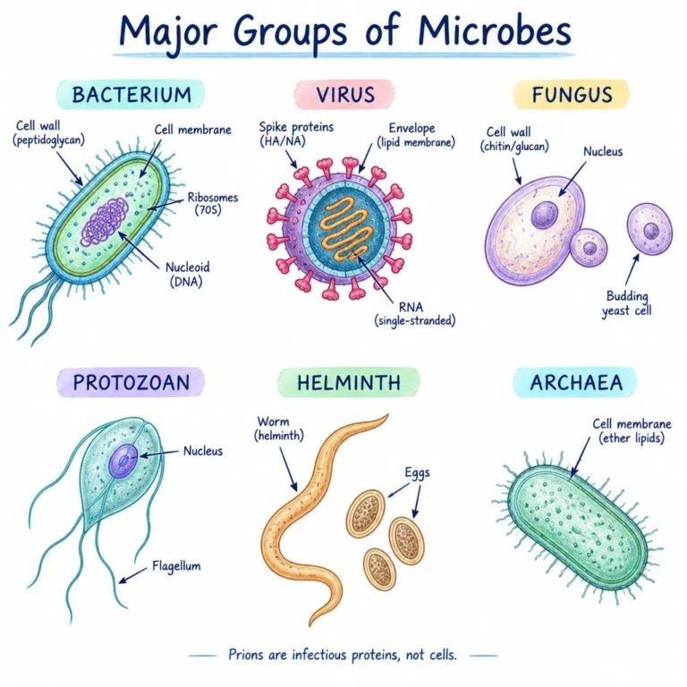
Bacteria vs Viruses vs Fungi: What is the main Difference?
Bacteria are cells that can reproduce independently in suitable environments. Viruses must use host cellular machinery to make new virus particles. Fungi are eukaryotic organisms that include yeasts and multicellular molds. Therefore antibacterial antibiotics generally do not treat viruses or fungi; antimicrobial drug classes differ by the organism and condition.
What is a Pathogen?
A pathogen is an organism capable of causing disease. Pathogenicity refers to that capability; virulence describes the degree of damage or severity under relevant circumstances. Opportunistic pathogens are more likely to cause disease when host defenses or normal barriers are weakened. The mere presence of a microorganism does not establish an active infection.
3. Prokaryotic vs Eukaryotic cells: Nursing Microbiology Comparison
Direct answer for exams: Prokaryotic cells do not have a membrane bound nucleus; eukaryotic cells do. The distinction also involves the organisation of DNA, ribosomes and internal organelles. Human cells and fungal cells are eukaryotic, while bacteria are prokaryotic. Viruses are not cells and belong to neither group.
What do Prokaryotic and Eukaryotic cells have in common?
Both have a plasma membrane, genetic material, ribosomes and cytoplasm, and both carry out biochemical reactions. Therefore, the statement that “only eukaryotes have DNA or ribosomes” is incorrect. The important differences concern how those structures are organised and, in many cases, their molecular characteristics.
Are Bacteria Prokaryotic or Eukaryotic?
Bacteria are prokaryotic organisms. Their DNA usually occupies a nucleoid rather than a membrane bound nucleus. They may also contain plasmids, and many have a peptidoglycan cell wall. Human cells are eukaryotic; the lack of peptidoglycan in human cells helps explain why some antibacterial medicines can act selectively against bacterial cell-wall synthesis. Drug selectivity is never absolute, so medicines can still cause adverse effects.
How does a Fungal cell differ from a Bacterial Cell?
A fungal cell has a nucleus and membrane bound organelles. Many fungi possess cell walls rich in chitin and glucans, whereas many bacteria have peptidoglycan. This distinction matters because antifungal and antibacterial medicines target different biological features. A yeast such as Candida is not a bacterium even though it may appear in a clinical specimen.
Common examination mistake: Prokaryotic does not mean “always disease-causing,” and eukaryotic does not mean “multicellular.” Many fungi are unicellular yeasts and many prokaryotes are harmless environmental organisms.
Prokaryotic cells lack a membrane bound nucleus and include bacteria and archaea. Eukaryotic cells have a membrane bound nucleus and organelles; human cells, fungi and protozoa are eukaryotic. Viruses are neither prokaryotic nor eukaryotic cells.
| Feature | Prokaryotic cells | Eukaryotic cells |
|---|---|---|
| Nucleus | No membrane bound nucleus; DNA in nucleoid region | Membrane-bound nucleus |
| Genetic material | Often a circular chromosome in bacteria; exceptions exist | Usually multiple linear chromosomes in the nucleus |
| Membrane-bound organelles | Absent | Present, e.g., mitochondria and endoplasmic reticulum |
| Ribosomes | Typically 70S in bacteria | Typically 80S cytoplasmic ribosomes |
| Cell wall | Most bacteria have peptidoglycan; some exceptions | Animals lack cell walls; fungi have walls mainly containing chitin and glucans |
| Typical size | Often smaller, but sizes overlap | Often larger, but not always |
| Examples | E. coli, S. aureus | Human cells, Candida, Plasmodium |
Easy memory point: Prokaryote: no membrane bound nucleus; Eukaryote: true membrane bound nucleus. The distinction also helps explain why some antibacterial drugs can target bacterial structures that differ from human cells.
Do all bacteria have a cell wall?
No. Many bacteria have a peptidoglycan wall, but Mycoplasma species lack a conventional cell wall. Do not make absolute statements that every bacterium stains reliably by the Gram method or responds to antibiotics that target bacterial cell walls.
4. Bacterial Cell Structure, Shapes and Arrangements
Search-intent summary: Bacterial morphology means the observable form and arrangement of bacterial cells. The three basic shapes taught in introductory microbiology are cocci (round), bacilli (rod-shaped) and spiral forms. More specific terms include vibrio (curved rod), spirillum (relatively rigid spiral) and spirochete (flexible spiral). Shape is only one part of identification.
What is the difference between Cocci, Bacilli and Spirilla?
Cocci are rounded cells, bacilli are rods, and spirilla have a spiral form. The term coccobacilli describes short rods that resemble an intermediate shape. The words streptococci and staphylococci refer to arrangements often described as chains and clusters respectively; the shape and arrangement should be described separately.
How do Diplococci, Streptococci and Staphylococci differ?
| Arrangement | Appearance | Revision cue |
|---|---|---|
| Diplococci | Two round cells together | Diplo = pair |
| Streptococci | Round cells arranged in chains | Strepto = chain |
| Staphylococci | Irregular clusters of round cells | Staphylo = cluster |
| Diplobacilli | Two rod-shaped cells together | Pair of rods |
| Streptobacilli | Chains of rod-shaped cells | Chain of rods |
Nursing interpretation: “Gram positive cocci in clusters” reports a laboratory staining and morphology pattern. It does not, without further tests, prove the presence of a particular Staphylococcus species or establish that a collected specimen represents active infection.
Parts of a Bacterial cell and their Functions
| Structure | Function | Clinical relevance |
|---|---|---|
| Cell membrane | Selective barrier for transport and cellular processes | Maintains cell integrity and physiology |
| Peptidoglycan cell wall | Helps determine shape and withstand osmotic pressure | Underlying basis of Gram response and target of some antibiotics |
| Capsule / glycocalyx | Outer covering in some species; helps adhesion and survival | May influence immune evasion and biofilm formation |
| Cytoplasm | Contains enzymes, ribosomes and metabolic components | Site of many biochemical reactions |
| Nucleoid | Region containing bacterial chromosome | Stores genetic information |
| Plasmid | Additional DNA molecule in some bacteria | May carry certain antimicrobial-resistance genes |
| Ribosomes | Protein synthesis | Different from human cytoplasmic ribosomes; drug-target relevance |
| Flagella | Motility in certain bacteria | Contributes to movement, not present in all species |
| Pili / fimbriae | Attachment; some pili participate in genetic exchange | May help colonization or transfer genetic material |
| Endospores | Dormant, highly resistant survival structures produced by some bacteria | Important to reprocessing and infection prevention concepts |
Bacterial Shapes: Cocci, Bacilli, Vibrios, Spirilla and Spirochetes
| Shape | Appearance | Examples / arrangement |
|---|---|---|
| Cocci | Round or roughly spherical | Pairs (diplococci), chains (streptococci), clusters (staphylococci) |
| Bacilli | Rod-shaped | Single rods, pairs or chains; E. coli is a bacillus |
| Vibrios | Curved, comma-shaped rods | Vibrio species |
| Spirilla | Relatively rigid spiral-shaped bacteria | Spirillum-type shapes |
| Spirochetes | Thin, flexible corkscrew-shaped bacteria | Treponema, Borrelia |
Exam distinction: Shape describes a bacterial cell’s appearance; arrangement describes how cells remain grouped after division. A single organism may show shape variation, and microscopy alone usually cannot identify an exact bacterial species.
What are Bacterial Endospores?
Endospores are tough dormant structures formed by certain bacterial groups, including some Bacillus and Clostridium-related organisms. They withstand environmental stresses that kill many vegetative bacterial cells. An endospore is not the same thing as a reproductive fungal spore, and ordinary cleaning or low-level disinfection is not equivalent to validated sterilization.
5. Gram Positive vs Gram Negative Bacteria and Gram Staining
Fast answer: In a typical Gram stain, Gram positive bacteria retain the violet primary dye and look purple; Gram negative bacteria are decolorized and take up the pink/red counterstain. The result reflects cell-envelope properties, especially the peptidoglycan layer and outer membrane. It does not measure the organism’s virulence or determine an antibiotic prescription.
What is the principle of Gram staining?
Gram staining differentiates many bacteria according to how their cell envelopes respond to sequential staining and decolorization. Crystal violet stains the cells, iodine helps stabilise the crystal-violet complex, a decolorizer removes it more readily from typical Gram negative cells, and safranin makes those decolorized cells visible in pink or red. A suitable specimen, correct staining technique and microscopic interpretation are necessary to obtain a useful result.
Why do Gram Positive bacteria appear purple and Gram Negative bacteria appear pink?
The thick peptidoglycan envelope of typical Gram positive bacteria helps retain the purple dye complex after decolorization. Typical Gram negative bacteria have a relatively thin peptidoglycan layer and an outer membrane; the decolorization step removes the purple complex sufficiently for the counterstain to be seen. This is a useful structural explanation, although real organisms may be Gram-variable or poorly visualized.
What is the difference between Gram Staining, Acid fast staining and culture?
| Method | Primary purpose | What it cannot establish alone |
|---|---|---|
| Gram stain | Describe cell-wall staining group and bacterial morphology | Exact species, active infection, drug susceptibility |
| Acid-fast stain | Help identify acid-fast organisms with distinctive envelope properties | Species and clinical significance without further testing |
| Culture | Grow recoverable organisms in a suitable medium | Whether a positive result is contamination, carriage or disease without context |
| Susceptibility testing | Assess an isolate’s response to selected antimicrobial agents in vitro | Patient response without considering exposure, infection site and other factors |
Common Gram stain interpretation mistakes
- “Purple means more dangerous”: incorrect. Both groups contain pathogens and harmless organisms.
- “A Gram stain tells us exactly which antibiotic will work”: incorrect; antimicrobial susceptibility needs its own assessment.
- “All bacteria stain reliably by Gram stain”: incorrect; some organisms need specialized methods.
- “A positive stain proves an infection”: incorrect; specimen quality and patient findings matter.
Exam-ready result wording: A lab may report Gram negative rods, Gram positive cocci in pairs or Gram positive cocci in clusters. Each describes the observed stain and morphology, not a final clinical diagnosis.
Gram staining is a differential staining method that groups many bacteria according to how their cell envelope retains dyes. It is a fast laboratory clue, not a definitive pathogen identification or treatment decision.
| Feature | Gram positive bacteria | Gram negative bacteria |
|---|---|---|
| Stain appearance | Usually purple/violet | Usually pink/red after counterstaining |
| Peptidoglycan | Relatively thick layer | Relatively thin layer |
| Outer membrane | Typically absent | Present in classical Gram negative bacteria |
| Lipopolysaccharide (LPS) | Absent as a typical outer-membrane feature | Present in many Gram negative outer membranes |
| Examples | Staphylococcus, Streptococcus | E. coli, Klebsiella |
| Important caution | Not all Gram positive organisms are harmless or susceptible to one antibiotic | Not all Gram negative organisms cause the same illness or share resistance patterns |
Gram staining steps and their purpose
- Crystal violet: the primary stain colors cells purple.
- Gram iodine: acts as a mordant and helps form dye complexes.
- Decolorization: alcohol or acetone removes the primary dye more readily from typical Gram negative cells; this step is technique-sensitive.
- Safranin: counterstains decolorized cells pink/red while Gram positive cells generally remain purple.
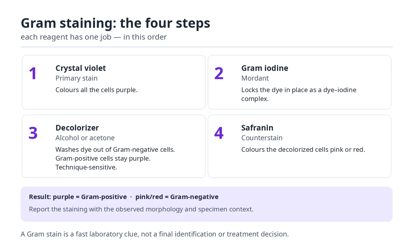
How to interpret a Gram stain: report the staining response with observed morphology and specimen context. For example,, Gram positive cocci in clusters. The final diagnosis may require culture, molecular identification, susceptibility testing and clinical assessment. Some bacteria are Gram-variable or unsuitable for reliable Gram staining.
Gram Positive versus Gram Negative cell-wall concept
Quick visual summary: Gram positive → thick peptidoglycan → retains crystal violet (purple); Gram negative → thinner peptidoglycan plus outer membrane → decolorized and counterstained pink. This is a model of typical patterns, not a complete classification of all bacteria.
Nursing application: A Gram stain on a relevant clinical specimen can provide preliminary clues to the care team. Nurses support accurate specimen collection and safe transport, monitor the patient and communicate concerning findings; antibiotic selection is based on authorised clinical decisions.
6. Bacterial Reproduction, Binary Fission and the Bacterial growth curve
Definition students search: The bacterial growth curve is a graph that describes how a bacterial population changes over time in a closed culture. The four stages are lag phase, log (exponential) phase, stationary phase and death (decline) phase. The horizontal axis shows time and the vertical axis usually shows the logarithm of viable bacterial count. This is a laboratory population graph, not a chart of a patient’s recovery.
Why is the log phase called exponential growth?
During suitable conditions, repeated binary fission produces an increase that is proportional to the number of cells already present. When the doubling time remains constant, each new generation doubles the population: 1, 2, 4, 8, 16 and so on. On a conventional linear axis the rise curves upward; on a logarithmic y-axis, an ideal exponential phase appears approximately as a straight rising line. That is why students must label graph axes carefully.
How to calculate bacterial population after several generations
For a simple textbook problem, use N = N0 × 2n, where N0 is the initial cell count and n is the number of doublings. If 200 bacteria double three times under ideal conditions, the expected count is 200 × 2³ = 1,600 cells. To find n, divide the elapsed growth time by the generation time when that time is constant. This mathematical model assumes sufficient resources and does not apply unchanged to real infections.
Bacterial growth curve questions examiners commonly ask
- Where do cells adapt to the environment? Lag phase.
- Where is multiplication fastest? Log phase, under the specified growth conditions.
- Where do nutrient exhaustion and waste accumulation matter? Especially as the culture enters stationary and decline phases.
- Do all bacteria grow at the same rate? No. Growth depends on the species and environmental conditions.
- Does the curve demonstrate resistance? No; resistance requires separate microbiological evidence.
Nursing connection: The growth curve builds understanding of culture-based testing and why specimen timing, environmental conditions and microbial physiology matter. It does not provide a safe method for predicting infection severity, choosing medicines or timing patient isolation.
How do bacteria reproduce?
Most bacteria reproduce through binary fission. A cell replicates its DNA, grows and divides into two daughter cells. Under suitable conditions repeated doubling can increase cell numbers quickly, although real growth rates vary substantially by organism and environment.
Four phases of the bacterial growth curve
In a closed batch culture, the log of viable bacterial cell count against time often forms a curve with four phases. This teaching model describes growth in a defined culture system; it must not be used as a direct timeline for a patient’s infection.
| Phase | What happens | Common exam answer |
|---|---|---|
| Lag phase | Cells adjust, synthesize needed components and may repair injury; little net population growth | Adaptation |
| Log (exponential) phase | Cells divide rapidly; the population grows exponentially | Active multiplication |
| Stationary phase | Nutrient limitation and waste accumulation produce an approximate balance between cell division and death | Growth plateaus |
| Death (decline) phase | Viable cell count decreases as death exceeds division | Population declines |
Text diagram: Lag (nearly level) → Log (steep rise) → Stationary (plateau) → Decline (downward slope). When sketching the graph, label the horizontal axis time and the vertical axis log of viable bacterial cells.
What is bacterial generation time?
Generation or doubling time is the time required for a bacterial population to double under specified conditions. If a hypothetical population starts at 100 cells and doubles twice, the expected number is 100 × 2² = 400. This calculation assumes no cell loss or environmental limitation; it is not a prediction for patient infection.
What factors affect bacterial growth?
- Nutrients and available energy sources
- Temperature and pH
- Oxygen requirements (aerobic, anaerobic and facultative organisms)
- Water availability and osmotic conditions
- Competition, waste accumulation and antimicrobial stress
- Surface attachment and biofilm formation
What is a biofilm?
A biofilm is a community of microorganisms attached to a surface and embedded in an extracellular matrix. Biofilms can develop on surfaces including certain medical devices. They may be harder to remove and can have altered susceptibility to antimicrobials. Prevention, device assessment and correct device-care protocols are essential nursing connections.
7. Normal Flora (Normal Microbiota): Examples, Functions, Body Sites and Nursing Importance
What is normal flora in microbiology?
Normal flora is the traditional term for microorganisms that normally live on or in certain parts of the human body without necessarily causing disease. The preferred scientific term is normal microbiota. These communities include bacteria and fungi and, more broadly, can include other microorganisms. The microbiome may refer to the microorganisms together with their genes and surrounding environment; the terms are related but are not perfectly interchangeable.
For example, Staphylococcus epidermidis commonly lives on healthy skin, various Streptococcus species inhabit the mouth, and large communities of anaerobic bacteria occupy the colon. An organism may be harmless at its usual body site but harmful when it reaches a wound, bloodstream, urinary tract or another vulnerable site. Therefore, “normal” does not mean sterile, harmless in every circumstance, or identical in every person.
Which parts of the body contain normal flora? Examples by body site
The skin, oral cavity, nose, intestinal tract and genital mucosa provide different environments for microbial life. Moisture, oxygen, nutrients, acidity, hormones, hygiene and host defenses influence which organisms are common. These are illustrative examples, not a universal organism list; individual microbiota vary considerably.
| Body site | Examples of normal or commonly carried microorganisms | What students should understand |
|---|---|---|
| Skin | Staphylococcus epidermidis, Corynebacterium species, Cutibacterium acnes | These organisms occupy skin niches and interact with the skin barrier. Skin flora can contaminate specimens or contribute to infection if introduced through invasive devices or damaged skin. |
| Anterior nose (nasal carriage) | Various staphylococci and corynebacteria; some individuals also carry Staphylococcus aureus | Carriage of S. aureus can occur without illness, but the species has important disease-causing potential. Carriage is not equivalent to an active infection. |
| Mouth and upper throat | Oral streptococci such as Streptococcus salivarius, plus Actinomyces, Veillonella and other anaerobes | Microbes live in saliva, dental plaque and gingival crevices. Some contribute to oral health, while shifts in oral communities and plaque conditions can be associated with dental caries or periodontal disease. |
| Large intestine (colon) | Bacteroides, Bifidobacterium and many other anaerobes; Escherichia coli may be present as a smaller component | Gut communities help process food components and resist colonization by some pathogens. Intestinal organisms can become opportunistic pathogens outside the bowel, such as E. coli in some urinary tract infections. |
| Vagina | Lactobacillus species commonly predominate in many reproductive-age individuals; mixed communities also occur, and Candida may be carried without symptoms | Lactobacilli often help maintain an acidic environment. Composition varies with age, hormonal state and individuals; a laboratory finding must be interpreted with symptoms and the specific condition. |
| Distal urethra and surrounding skin | Mixed skin and mucosal organisms, including some coagulase-negative staphylococci and corynebacteria | Microbes at the opening can enter a urine specimen during collection. This does not mean every positive urine culture indicates a urinary tract infection. |
Important distinction: The location of a microorganism matters. Skin and mucosal surfaces normally host communities; blood and deep tissues are not expected to have established normal bacterial flora. A blood-culture result may represent true bloodstream infection or specimen contamination and requires prompt, appropriate clinical interpretation.
What are the main functions of normal flora?
1. Protection against harmful microorganisms (colonization resistance)
Resident microorganisms compete with newcomers for attachment sites and nutrients. Some also produce substances that discourage the growth of competing microbes or help maintain local conditions that do not favor certain pathogens. This protection is called colonization resistance; it reduces risk but does not make a person immune to infection. A useful nursing example is the intestinal microbiota, whose disruption can make it easier for Clostridioides difficile to multiply.
2. Digestion and microbial metabolism
Gut microorganisms break down some dietary components that human enzymes cannot digest efficiently, including certain fibers. Their metabolic products include short-chain fatty acids, such as acetate, propionate and butyrate, which contribute to normal intestinal physiology. Some gut organisms also synthesize vitamins, including forms of vitamin K and certain B vitamins; the amount actually available to the host varies and should not be treated as a substitute for adequate nutrition.
3. Interaction with immune defenses
The microbiota communicates with epithelial barriers and immune cells, helping shape how the body responds to ordinary microbial exposure. In a healthy state, these interactions contribute to barrier integrity and the balance between defense and unnecessary inflammation. The microbiota works alongside innate and adaptive immunity; it does not replace white blood cells, antibodies or vaccination.
4. Support for local body environments
Different body sites have distinct chemical conditions. For example, certain vaginal lactobacilli produce lactic acid, helping maintain an acidic environment that can discourage growth of some other organisms. On the skin, resident microbes interact with sebum, moisture and the skin barrier. These mechanisms help explain why normal flora varies across the body instead of forming one identical community everywhere.
Resident flora versus transient flora: how are they different?
Resident flora consists of microorganisms relatively well adapted to a body site and commonly present over time. Transient flora refers to organisms acquired temporarily through contact with other people, patient-care environments, surfaces, food or equipment. A transient organism may disappear or may colonize if conditions allow; duration is not a fixed rule for every microbe.
| Feature | Resident flora | Transient flora |
|---|---|---|
| Usual pattern | Established at a particular site | Acquired for a shorter period |
| Examples | S. epidermidis on skin; many colon bacteria | Organisms recently transferred onto hands after patient or surface contact |
| Potential benefit or risk | May protect the host but can cause opportunistic infections | May be harmless or may be transmissible pathogens |
| Nursing significance | Protect skin integrity and apply aseptic technique for invasive care | Perform hand hygiene at the recommended moments and prevent cross-transmission |
Practical example: A nurse may normally have resident skin flora on their hands and acquire transient organisms after touching a contaminated bedside surface. Gloves do not replace hand hygiene, and neither “resident” nor “transient” means that a microorganism is always safe or always harmful.
Normal flora, colonization, infection and contamination: what is the difference?
| Term | Meaning | Clinical example |
|---|---|---|
| Normal microbiota | Microorganisms normally associated with a body site | S. epidermidis on intact skin |
| Colonization | Microorganisms are present and may multiply without causing apparent disease | Nasal carriage of S. aureus without symptoms |
| Infection | A microorganism establishes a host interaction that can cause tissue damage or a disease process; some infections are asymptomatic | Symptomatic urinary infection supported by assessment and appropriate testing |
| Specimen contamination | Microorganisms enter the sample unintentionally during collection, storage or handling | Skin organisms introduced into a blood-culture bottle during collection |
A positive culture alone is not always proof of symptomatic infection. Clinical signs, the type and quality of the specimen, organism identity, collection method and patient context all matter. For example, asymptomatic bacteriuria means bacteria are found in urine in a person without symptoms attributable to a urinary tract infection. In many groups, treatment is not recommended; important exceptions include pregnancy and certain invasive urologic procedures. Nurses should follow the clinician’s orders and applicable guidelines rather than independently prescribing or assuming all bacteriuria needs antibiotics.
How can normal flora become harmful? Opportunistic infection examples
Opportunistic pathogens cause disease more readily when they enter an unsuitable site or when host defenses are impaired. A microorganism’s effect depends on the site, host and circumstances rather than its name alone.
- Escherichia coli: It can live in the intestine without causing illness there. Certain strains may cause urinary tract infection after reaching the urinary tract, particularly when conditions favor ascent and multiplication. This is why specimen collection and symptom assessment are important.
- Staphylococcus epidermidis: Common skin carriage is normally harmless, but the organism can cause a clinically important infection in association with some intravenous catheters, prosthetic devices or other implanted materials. It can also contaminate blood cultures, so a laboratory result needs contextual interpretation.
- Candida species: Yeasts may be present on mucosal sites without causing symptoms. Under some circumstances, including changes in local conditions or impaired host defenses, overgrowth may contribute to mucosal candidiasis; invasive disease is a distinct serious condition.
- Bacteroides species: Many are established intestinal anaerobes but can be involved in abscesses or other infections if intestinal contents enter normally protected tissues, such as after bowel perforation.
What is dysbiosis and what changes normal flora?
Dysbiosis means a change in the composition or functioning of microbial communities that is associated with disturbance of their usual ecological balance. It is a broad descriptive concept, not a diagnosis that can be made from a single organism or one universal “healthy microbiome” score. Diet, antibiotics, illness, age, hormones, environmental exposure and other factors can affect microbiota composition.
Example: antibiotics and C. difficile. Antibiotics can be essential for treating bacterial infections, but some also reduce helpful gut bacterial populations. This can weaken colonization resistance, allowing C. difficile to multiply and cause toxin-mediated diarrhea in susceptible patients. Nurses should monitor for new unexplained diarrhea, communicate concerns, use indicated precautions and follow institutional protocols. This does not mean that every episode of diarrhea following antibiotics is C. difficile infection.
Why is normal flora important in nursing practice?
- Interpret specimens carefully: help collect properly labelled, appropriately timed specimens using the ordered procedure. Recognize that an organism on the skin or mucosa may represent carriage, contamination or an infection depending on the sample and clinical findings.
- Prevent cross-transmission: follow Standard Precautions, the recommended hand-hygiene moments, appropriate PPE and equipment decontamination. The presence of normal flora never removes infection-control responsibilities.
- Protect invasive-device sites: use approved aseptic and device-care protocols because skin organisms can sometimes enter tissues or the bloodstream through a catheter or other device.
- Support antimicrobial stewardship: communicate symptoms and specimen findings accurately. Antibiotics can affect both pathogens and beneficial bacteria, so unnecessary exposure should be avoided according to prescribing guidance.
- Recognize changes in vulnerable patients: compromised skin, recent antibiotics, invasive devices and altered immune defenses may increase the importance of normally carried organisms.
- Explain results without alarming patients: finding bacteria on a swab or in a specimen does not always mean a dangerous infection. Explain that the clinical team interprets the result alongside symptoms and history.
Worked clinical examples: apply the concept
Case A — Positive nasal swab: An adult patient has a screening swab positive for S. aureus, but no local symptoms. This finding may represent colonization, not an active nasal infection. The nurse follows the facility’s screening, infection-control and communication procedures without assuming antibiotic treatment is required solely from the screening result.
Case B — Urine culture without urinary symptoms: An adult has bacteria reported in urine but no symptoms attributable to a urinary tract infection. The result may fit asymptomatic bacteriuria; management depends on the patient group and applicable guidelines. The nurse confirms relevant symptoms, specimen quality and patient factors, then communicates with the responsible clinician.
Case C — Skin organism in a blood culture: A culture grows a coagulase-negative staphylococcus. Because these organisms commonly live on skin, specimen contamination is one possibility, but a true infection is also possible, particularly with devices or compatible patient findings. The nurse must not dismiss or independently diagnose the result; the responsible team assesses the complete clinical picture.
Normal flora: important examination questions with explained answers
Q1. Define normal flora and give five examples.
Normal flora (normal microbiota) consists of microorganisms normally associated with particular human body sites. Examples include Staphylococcus epidermidis and Cutibacterium acnes on skin, Streptococcus salivarius in the mouth, Bacteroides species in the colon, and Lactobacillus species in many vaginal communities. An example is only meaningful when its usual body site and potential clinical role are also considered.
Q2. Explain four functions of normal microbiota.
Normal microbiota can limit pathogen establishment by competing for nutrients and attachment sites; help metabolize dietary substrates and produce useful metabolites; interact with immune defenses and barrier tissues; and support local conditions such as vaginal acidity. These functions vary by body site and do not guarantee freedom from disease.
Q3. Differentiate normal flora, colonization and infection with an example.
Normal flora describes communities typically present at body sites; colonization means an organism is present without necessarily causing disease; and infection means a disease-related host–microorganism interaction has developed, although some infections may be asymptomatic. S. aureus carriage in the nose may be colonization, while a confirmed S. aureus wound infection involves a different clinical situation. Interpretation requires findings beyond organism detection alone.
Q4. Why might antibiotics increase the risk of some infections?
Antibiotics intended to treat pathogens may also disturb helpful gut microorganisms. When colonization resistance declines, certain organisms, including C. difficile, can multiply in susceptible individuals and cause disease. This illustrates why appropriate antimicrobial use, clinical monitoring and infection prevention are linked.
Reliable references for further study
Review the NCBI Bookshelf chapter on normal flora, the University of Iowa’s body-site organism reference, CDC guidance on the gut microbiome and C. difficile, and the IDSA asymptomatic bacteriuria guideline. These explain the educational principles; local clinical protocols and clinician assessment govern care.
8. Chain of infection, modes of transmission and stages of infection
Answer in one sentence: The chain of infection is a model with six links: infectious agent, reservoir, portal of exit, mode of transmission, portal of entry and susceptible host. Together, these explain how a pathogen can spread. Infection prevention aims to interrupt one or more of those links. Students often search for the chain of infection in nursing, its correct order and a clinical example; the same concept applies in hospitals, clinics and communities.
Chain of infection example: contaminated hands and a wound
Imagine a microorganism present in wound drainage (agent and reservoir). The drainage leaves the wound (exit), contaminates a glove or surface, and is then transferred by contact (transmission). If transferred to another vulnerable wound (entry), it may reach a susceptible patient. Nurses can interrupt this sequence through hand hygiene, glove changes between tasks, appropriate wound protection, equipment cleaning and correct waste handling. The example illustrates a possible route, not evidence that every contaminated surface will cause an infection.
Chain of infection example: respiratory illness
A respiratory pathogen may leave an infected person in exhaled respiratory particles and reach another person’s mucous membranes or airways. Source control, appropriate patient placement, ventilation and risk-based respiratory protection can interrupt relevant links. The precise combination depends on the organism and the setting. Do not assume that all respiratory pathogens require identical room placement or PPE.
Reservoir vs source vs portal of exit: what is the difference?
A reservoir is the environment or host in which a microorganism normally lives and may multiply. A source is the immediate person or object from which an infection is acquired; it may differ from the ultimate reservoir. The portal of exit is the route through which the organism leaves that reservoir or source. For exam answers, name the links separately instead of using reservoir and transmission interchangeably.
Chain of infection vs infection cycle vs stages of infection
The phrase infection cycle is sometimes used informally for transmission, but it can also refer to an organism’s life cycle. By contrast, the stages of infectious disease describe the time course of illness in a person. When answering an examination question, use the terminology in the question and state whether you are explaining transmission between hosts or progression within a host.
What are the six links in the chain of infection?
- Infectious agent: the microorganism capable of causing disease.
- Reservoir: where the organism lives or survives, such as a person, equipment or environment.
- Portal of exit: how the agent leaves, for example via respiratory secretions or blood.
- Mode of transmission: how it reaches another host through contact, droplets, inhalation of infectious aerosols, vehicles or vectors as appropriate.
- Portal of entry: a route into the host, such as mucous membranes or broken skin.
- Susceptible host: someone at risk because of relevant host factors.

How can a nurse break the chain of infection?
| Link to interrupt | Example of prevention |
|---|---|
| Agent | Appropriate reprocessing and infection-control practices, including prescribed therapy where indicated |
| Reservoir | Environmental cleaning, safe equipment handling and relevant source control |
| Portal of exit | Respiratory hygiene, safe containment of body fluids and wound drainage |
| Transmission | Hand hygiene, PPE chosen according to the risk and transmission-specific precautions |
| Portal of entry | Aseptic technique and protecting invasive-device entry sites |
| Susceptible host | Vaccination where indicated, protection of skin integrity and reduction of avoidable exposures |
Contact, droplet and airborne transmission
Contact transmission may involve direct contact or contaminated objects. Droplet transmission involves respiratory droplets and relevant close exposures. Airborne transmission involves infectious particles that can remain suspended and be inhaled. Organism-specific guidance determines which precautions apply; some infections can involve more than one route.
What are the stages of an infectious disease?
- Incubation: time between exposure and onset of symptoms, if symptoms develop.
- Prodromal stage: early, often non-specific symptoms in diseases that have this stage.
- Illness stage: more characteristic manifestations may be present.
- Decline: manifestations diminish as the illness resolves or responds to treatment.
- Convalescence: recovery toward previous function.
Do not confuse: the chain of infection explains transmission between hosts; the stages of infection describe a possible time course within an individual. Not every infection follows every stage or produces symptoms.
Healthcare-associated infections (HAIs)
A healthcare-associated infection is acquired in association with healthcare delivery, rather than being present or incubating at the time care begins according to the applicable surveillance definition. Common prevention priorities include device care, aseptic practice, environmental cleaning, hand hygiene and evidence-based surveillance. Different infections have different prevention bundles and diagnostic criteria.
9. Infection prevention for nurses: standard precautions and isolation
Direct answer: Standard Precautions are the baseline practices applied to all patient care. Transmission-Based Precautions are additional measures used when a suspected or confirmed organism or syndrome presents a transmission risk not adequately controlled by Standard Precautions alone. The three main categories are contact, droplet and airborne precautions, and more than one category can apply.
What are the main components of Standard Precautions?
Standard Precautions include hand hygiene; exposure-PPE chosen according to the risk; respiratory hygiene; safe injection and sharps practices; appropriate environmental cleaning; and safe handling, cleaning, disinfection or sterilization of equipment. They do not mean every patient needs every item of PPE. A nurse chooses protection based on anticipated contact with blood, body fluids, respiratory particles or contaminated surfaces and the current facility guidance.
Contact precautions: what PPE is generally used?
For a patient requiring Contact Precautions, guidance commonly includes a gown and gloves for relevant patient and environmental contact, along with proper placement, equipment and cleaning. Standard Precautions still apply. Exact procedures depend on the organism, facility, healthcare setting and current policy. Some organisms require additional measures or particular hand-hygiene methods.
Droplet precautions: is a surgical mask enough?
Droplet Precautions commonly require appropriate medical masking when entering the patient care space and actions such as patient source control and suitable room placement. Other protection may be needed following risk assessment and current guidance. Do not use a generic mask rule as a substitute for the facility’s disease-specific requirements or for airborne protection when indicated.
Airborne precautions: why is a respirator different from a mask?
Airborne Precautions generally require a suitable airborne infection isolation room and an appropriately fit-tested respirator such as an N95 or higher-level approved device for healthcare personnel, as determined by local regulation and policy. A surgical mask is not equivalent to a fit-tested respirator. The correct approach also depends on facility capabilities and emergency transfer protocols.
Which precaution is needed for a particular organism?
Rather than memorizing a single fixed disease-to-PPE table, use current organism-specific guidance. For example, tuberculosis is a classic example requiring airborne-focused precautions; other infections may call for contact, droplet or combined precautions. An uncertain diagnosis does not mean precautions must always await laboratory confirmation. Contact the clinical or infection prevention team when the patient’s presentation suggests a transmissible infection.
Safety reference: CDC Standard Precautions and CDC Transmission-Based Precautions. These pages help clarify the principles but do not replace the approved local protocol.
Standard Precautions vs Transmission-Based Precautions
| Precautions | When used | Main measures |
|---|---|---|
| Standard Precautions | For every patient, regardless of diagnosis | Hand hygiene, PPE chosen according to the risk, respiratory hygiene, injection safety, equipment reprocessing and environmental cleaning |
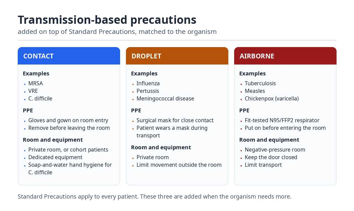
| Contact Precautions | In addition to Standard Precautions for relevant contact-transmitted organisms | Gloves and gown on room entry, removed before leaving; private room or cohorting; dedicated equipment. Examples: MRSA, VRE, C. difficile (soap-and-water hand hygiene for C. difficile) |
| Droplet Precautions | In addition to Standard Precautions for relevant droplet-spread infections | Surgical mask for close contact; patient wears a mask during transport; private room. Examples: influenza, pertussis, meningococcal disease |
| Airborne Precautions | In addition to Standard Precautions for relevant airborne infections | Fit-tested N95/FFP2 respirator before entering; negative-pressure room with door closed; limit transport. Examples: tuberculosis, measles, chickenpox |
Isolation is not a single universal protocol: PPE, room placement and duration depend on the suspected organism, risk assessment, available infrastructure and current institutional guidance. Follow the local infection prevention team and current CDC, WHO or relevant national guidance.
WHO Five Moments for Hand Hygiene
- Before touching a patient
- Before a clean or aseptic procedure
- After body-fluid exposure risk
- After touching a patient
- After touching patient surroundings
Gloves do not replace hand hygiene. Perform hand hygiene after glove removal and at all other indicated moments. Alcohol-based hand rub is preferred in many healthcare circumstances when hands are not visibly soiled; wash with soap and water when visibly soiled and when organism- or policy-specific directions require it.
Aseptic technique, PPE, sharps and environmental safety
- Medical asepsis: practices that reduce the number or spread of microorganisms; often called clean technique.
- Surgical asepsis: maintaining sterility for indicated procedures; requires a controlled sterile field and appropriate training.
- Personal protective equipment: gloves, gown, mask, respirator and eye protection selected through an exposure-risk assessment.
- Safe injections: single-use needles and syringes and correct medication handling prevent bloodborne and other transmission.
- Sharps: dispose of used sharps immediately in approved containers and follow local exposure-response pathways.
- Environmental cleaning: follow the surface, product, contact-time and frequency instructions approved for the area.
For additional supervised practical detail, see the existing WHO Five Moments nursing guide and aseptic wound care guide. These are related clinical skills, not prerequisites to understanding this single page Microbiology lesson.
10. Cleaning vs disinfection vs sterilization: definitions and nursing examples
| Method | Purpose | Important limitation |
|---|---|---|
| Cleaning | Physically removes soil, organic material and many microbes | Does not reliably kill all microbes; often required before disinfection/sterilization |
| Disinfection | Inactivates many or selected microorganisms on inanimate items according to the process and level | Not the same as sterilization; effectiveness depends on product and contact conditions |
| Antisepsis | Reduces microorganisms on living tissue with an approved antiseptic | Products must be suitable for skin or tissue, not general surfaces |
| Sterilization | Validated process intended to eliminate all viable microorganisms, including resistant bacterial spores | Requires suitable equipment, monitoring and manufacturer-approved processing |
| Asepsis | Practices intended to prevent contamination | Not a synonym for sterilization |
High-, intermediate- and low-level disinfection
Disinfectants differ in their permitted use and the range of organisms they inactivate. High-level disinfection can eliminate all microorganisms except high numbers of bacterial spores under specified conditions, while sterilization targets all viable microbial forms. The required method depends on the item and its intended contact with the patient; staff must follow validated facility reprocessing procedures.
Why is cleaning necessary before disinfection?
Organic material and soil can hinder effective contact between a disinfectant or sterilant and an item. Appropriate cleaning is therefore an essential part of many validated reprocessing workflows. Never replace device-specific instructions with a generic soak time or chemical concentration from an educational webpage.
11. Clinical microbiology laboratory: microscopy, culture media, tests and specimens
Quick comparison: A Gram stain describes microscopic appearance, a culture attempts to grow organisms, a culture and sensitivity test evaluates an isolate’s in vitro antimicrobial susceptibility, and PCR/NAAT detects selected nucleic-acid sequences. These tests answer different questions. The most useful method depends on the suspected organism, specimen and clinical reason for testing.
How do selective, differential and enriched culture media differ?
Selective media favour the growth of certain organisms over others by using ingredients that inhibit competing groups. Differential media show visible differences between organisms, often through indicator reactions. Enriched media contain additional nutrients that support more fastidious organisms. Categories are not mutually exclusive: one medium may be both selective and differential.
What does a culture and sensitivity (C&S) result tell us?
A culture result may identify an organism growing from an appropriate specimen. Antimicrobial susceptibility testing can report categories such as susceptible, intermediate/increased exposure or resistant according to the test system and standard used. These results do not independently determine whether the organism is causing symptoms or which treatment a specific patient should receive. Clinical interpretation includes test method, site of infection, available drug exposure, comorbidities and local stewardship advice.
Common specimen types in nursing microbiology
| Specimen | Common reason for microbiology testing | Important collection principle |
|---|---|---|
| Urine | Assess possible urinary infection in the right clinical context | Use the ordered collection method; prevent contamination; document relevant symptoms |
| Blood | Investigate possible bloodstream infection when clinically indicated | Follow approved aseptic identification, collection and transport procedures |
| Respiratory specimen | Investigate selected respiratory organisms | Use the correct specimen type, PPE and transport process |
| Wound specimen | Investigate suspected infection when appropriate | Follow the collection technique specified by the clinical and laboratory teams |
| Stool | Investigate gastrointestinal pathogens when indicated | Use proper containers, precautions and timely laboratory delivery |
Exam reasoning: A good answer does not stop at “send a culture.” Explain why patient identification, suitable specimen type, contamination prevention, correct labeling and transport matter to test accuracy.
Does PCR prove that an organism is alive?
Not necessarily. Molecular tests detect selected nucleic-acid targets; depending on the method, detectable material can persist even when organisms are no longer viable. The test’s intended use and performance characteristics determine how results should be interpreted.
Which methods help identify microorganisms?
| Method | What it shows | Nursing interpretation |
|---|---|---|
| Light microscopy and stains | Morphology, cellular features and preliminary staining patterns | Preliminary clues; not always a definitive identification |
| Culture | Growth of organisms in appropriate laboratory media | Can support organism identification; contamination/colonization must be considered |
| Culture and susceptibility testing | Which antimicrobials are active against an isolated organism under test conditions | Supports prescribed therapy; the clinical team interprets with infection site and patient factors |
| Antigen test | Presence of selected microbial component | Depends on organism, specimen, timing and test performance |
| PCR / NAAT | Target microbial nucleic acid | May detect genetic material even when viability is uncertain |
| Serology | A host antibody response to relevant antigens | Timing, immune status and test design influence interpretation |
Types of culture media: general-purpose, enriched, selective and differential
| Media category | Purpose | Example |
|---|---|---|
| General-purpose | Supports growth of many nonfastidious organisms | Nutrient agar |
| Enriched | Provides extra nutrients for more demanding organisms | Blood agar; chocolate agar |
| Selective | Discourages some groups while permitting others to grow | MacConkey agar selects many Gram negative enteric bacteria |
| Differential | Distinguishes certain metabolic or other characteristics | MacConkey can differentiate lactose fermenters; blood agar can show hemolysis |
Exam point: a medium may be both selective and differential. MacConkey agar is a common example. The color or appearance of colonies must be interpreted with the complete testing workflow; student notes do not substitute for a laboratory identification algorithm.
What is culture and sensitivity testing?
A culture may grow a microorganism from a specimen. Susceptibility testing then evaluates its in vitro response to selected antimicrobial agents under standardized conditions. The report is interpreted by the responsible clinician alongside infection site, organism identification, resistance mechanisms, patient factors and current treatment guidance.
Specimen collection: nursing responsibilities
- Confirm the correct patient, specimen type and authorised request.
- Explain the procedure and use the appropriate personal protective measures.
- Collect the ordered specimen using the current aseptic or clean collection protocol as applicable.
- Label specimens with required identifiers at the point of collection and document relevant timing.
- Ensure proper packaging, storage and prompt transport according to laboratory instructions.
- Escalate critical results or concerning patient changes through the clinical pathway.
Why is contamination important? Incorrect or contaminated collection can produce misleading results, unnecessary repeat tests or inappropriate treatment. Nurses help protect specimen validity by following the approved collection process, rather than improvising laboratory culture conditions.
What does a negative culture mean?
A negative result does not invariably exclude infection. Sampling timing, collection quality, previous medication, type of organism, specimen site and limits of the test all affect interpretation.
12. Immunity: innate vs adaptive; active vs passive; antigen vs antibody
Direct answers: Innate immunity responds through broadly acting barriers and recognition systems; adaptive immunity uses antigen-specific B- and T-cell responses and can develop immunologic memory. Active immunity results when the recipient’s immune system develops its own response, while passive immunity comes from receiving preformed antibodies. An antigen is something recognized by immune receptors; an antibody is a protein that binds an antigen.
What happens during the first and later encounters with an antigen?
For many adaptive immune responses, an initial exposure activates specific lymphocyte populations; some differentiate into effector cells, antibody-producing cells or memory cells. A later exposure may lead to a faster or stronger response because memory cells already exist. The timing and quality of memory differ by antigen, vaccine, infection and host factors. It is inaccurate to assume a previous infection always produces lifelong protection.
Innate vs adaptive immunity: example from a damaged skin barrier
Skin and mucous membranes help keep microbes out. When a barrier is disrupted, innate defenses such as local inflammatory responses and phagocytes become important. Adaptive immune responses may recognise specific antigens and generate targeted responses over time. These defenses interact continuously rather than operating as two fully separate systems.
Active versus passive immunity: natural and artificial examples
- Natural active: an individual develops a response after exposure to an infectious organism.
- Artificial active: vaccination stimulates an immune response.
- Natural passive: maternal antibodies transferred during pregnancy or through other physiological routes.
- Artificial passive: administration of preformed antibodies for a defined medical indication.
Exam misconception: Both active and passive immunity may produce protection, but receiving antibodies is not the same as stimulating the recipient to form durable immune memory. The duration of protection differs substantially across circumstances.
Antigen vs antibody in a laboratory test
An antigen test aims to detect a selected part or product of an organism. An antibody test generally looks for the body’s immune response against a specified antigen. A positive antibody finding cannot universally distinguish past infection from vaccination or current infection without knowing the assay, timing and clinical setting. Nursing students should distinguish what the laboratory target actually measures before trying to interpret a result.
Innate immunity vs adaptive immunity
| Feature | Innate immunity | Adaptive immunity |
|---|---|---|
| Response | Rapid, broadly pattern-based defense | Antigen-specific responses developed after exposure |
| Major components | Skin/mucosal barriers, phagocytes, complement, inflammatory responses | B lymphocytes, T lymphocytes and antigen-specific responses |
| Immunologic memory | No classic antigen-specific memory like adaptive immunity; some innate training phenomena exist | Typically develops memory following many exposures or vaccines |
| Examples | Intact skin, neutrophil response | Antibodies and memory T cells |
Active vs passive immunity
| Type | Where protection comes from | Duration / memory |
|---|---|---|
| Natural active | Immune response after an infection | May generate memory, variable durability |
| Artificial active | Vaccination stimulates immune response | May generate memory; boosters may be needed |
| Natural passive | Maternal antibodies transferred to fetus or infant | Temporary; no recipient-generated immune memory from the transfer |
| Artificial passive | Preformed antibodies administered as a medical intervention | Usually immediate but temporary |
Antigen vs antibody
An antigen is a molecular structure that can be recognized by an immune receptor or antibody. An antibody (immunoglobulin) is a protein made by activated B cells/plasma cells that binds a particular antigenic target. Antigens and antibodies are not interchangeable, and a positive antibody test may reflect past exposure, vaccination or another context depending on the assay.
Inflammation and the immune response
Inflammation recruits defensive mechanisms to respond to injury or infection. Classic local signs include redness, warmth, swelling, pain and sometimes reduced function, but not every infected patient displays all signs. Immune suppression, age and other host factors can change presentation.
Nursing application: Protect patients through recommended vaccination programmes, infection prevention and relevant monitoring. Use immune-test results only within their intended clinical interpretation.
13. Antibiotics, antimicrobials and antimicrobial resistance mechanisms
What is antimicrobial resistance? Antimicrobial resistance occurs when a microorganism can withstand an antimicrobial exposure that would ordinarily inhibit or kill susceptible organisms. Antibiotic resistance refers specifically to bacteria and antibacterial medicines. Resistance can be intrinsic to an organism or acquired through mutation or the exchange of genetic material. It is not the same as the patient becoming “immune to antibiotics.”
Intrinsic versus acquired antibiotic resistance
Intrinsic resistance reflects properties naturally present in a bacterial group. For example,, the absence of a drug’s target or an effective permeability barrier. Acquired resistance develops when a previously susceptible organism gains a resistance-associated change, sometimes through mutation or genetic transfer. Antimicrobial exposure can select for resistant populations; antibiotics do not purposefully teach bacteria how to resist them.
How do conjugation, transformation and transduction affect resistance?
Through conjugation, bacteria can transfer certain DNA elements through cell-to-cell contact. Through transformation, some bacteria can take up free DNA from their surroundings. Through transduction, bacterial viruses can move DNA between bacteria. Depending on the organisms and genes involved, these processes can spread resistance determinants. Not every resistance mechanism is carried on a plasmid, and not every organism uses all three routes.
Why is resistance different from an adverse drug reaction or treatment failure?
A drug may fail for reasons other than bacterial resistance: incorrect diagnosis, poor source control, drug exposure problems or host-related factors can contribute. Similarly, a patient’s adverse reaction does not imply that the organism is resistant. This distinction is important when monitoring prescribed therapy and documenting clinical response.
Antimicrobial stewardship in everyday nursing practice
- Verify the medication order and communicate allergies or relevant safety concerns.
- Collect ordered specimens correctly and at the time directed by the care team.
- Administer prescribed medicine according to institutional medication-safety policies.
- Monitor clinical response, report new symptoms and document observations.
- Use infection prevention to reduce the number of infections requiring treatment.
- Explain, without giving independent prescribing advice, why antibiotics are not a treatment for viral influenza or the common cold.
Practice question: A patient asks for antibiotics after a positive culture without symptoms. A strong answer describes the difference between colonization and disease, acknowledges exceptions, and explains that treatment decisions are based on the clinician’s assessment and applicable guidelines rather than on culture positivity alone.
What is an antimicrobial?
Antimicrobial is a broad term for an agent active against microorganisms. Antibiotics (antibacterials) act against susceptible bacteria; antiviral, antifungal and antiparasitic medications target other organisms or stages. Treatment is determined by the responsible clinician, not by organism shape or symptoms alone.
How do antibiotics generally work?
- Inhibiting bacterial cell-wall synthesis
- Interfering with bacterial protein synthesis
- Affecting nucleic-acid synthesis or function
- Disrupting certain microbial metabolic pathways
- Disrupting bacterial membrane function (for specific agents)
How do bacteria become resistant to antibiotics?
| Mechanism | Meaning | Illustrative example |
|---|---|---|
| Enzymatic inactivation | A bacterium makes an enzyme that deactivates some drugs | Some beta-lactamases |
| Target alteration | A bacterial target changes so a drug binds less effectively | Altered binding sites |
| Efflux pumps | Transport systems reduce drug accumulation inside cells | Drug efflux |
| Reduced permeability | Drug penetration into the cell is reduced | Changes affecting outer-membrane entry |
| Target protection / bypass | A bacterium protects the target or uses an alternative pathway | Alternative metabolic mechanisms |
| Biofilm-associated tolerance | Microbes in biofilms may be harder to eradicate | Device-associated biofilm situations |
Resistance is not the same as tolerance or persistence. Resistant organisms can grow despite defined antimicrobial concentrations; tolerant or persistent populations may survive exposure without necessarily meeting genetic resistance criteria. Mechanisms differ across drug–organism combinations.
Why does antibiotic stewardship matter?
Unnecessary or inappropriate antibiotic exposure can contribute to selection of resistance and cause avoidable harm. Nurses support antimicrobial stewardship by collecting correct specimens when ordered, administering prescribed medicines accurately, monitoring response and adverse effects, and communicating findings. Educating patients that antibiotics are not effective against viral illness is also important.
14. Common clinically relevant microorganisms: comparison table
The examples below illustrate important organism–condition relationships; they do not mean an organism always causes that disease or that a symptom identifies a single pathogen.
| Organism | Category | Relevant condition or context | Key nursing concept |
|---|---|---|---|
| Escherichia coli | Gram negative bacterium | Common cause of urinary tract infection | Interpret symptoms and specimen quality together |
| Staphylococcus aureus | Gram positive bacterium | Skin/wound infection, bloodstream infection | Precautions and correct wound care |
| Streptococcus pyogenes | Gram positive bacterium | Pharyngitis and some skin infections | Transmission prevention and assessment |
| Mycobacterium tuberculosis | Acid-fast bacterium | Tuberculosis | Appropriate airborne infection-control measures |
| Clostridioides difficile | Spore-forming bacterium | Certain antibiotic-associated diarrheal illnesses | Contact precautions and organism-specific hygiene policy |
| Influenza virus | Virus | Respiratory infection | Vaccination and recommended respiratory precautions |
| Hepatitis B virus | Virus | Bloodborne infection | Vaccination and sharps safety |
| Candida species | Fungus | Mucosal and invasive candidiasis | Host risk, correct identification |
| Plasmodium species | Protozoan parasite | Malaria | Travel/exposure history and diagnostic testing |
Examples of common clinical microorganisms: what nurses need to know
The summary table above is useful for revision, but each organism has a different reservoir, mode of transmission, clinical context and approach to laboratory investigation. The following examples connect the name of the organism to a realistic nursing situation. They are educational examples, not a method of identifying an infection from symptoms alone.
1. Escherichia coli — normal intestinal carriage and urinary infection
E. coli is a Gram-negative, rod-shaped bacterium and a familiar member of some human intestinal microbial communities. Although many strains are harmless in that location, certain strains may cause urinary tract infections, bloodstream infection or diarrheal disease. For a suspected UTI, nurses help collect an appropriate urine specimen, assess and document symptoms, and communicate concerning findings. A positive urine culture without attributable symptoms is not automatically a reason for antibiotic treatment; important patient-specific exceptions apply.
2. Staphylococcus aureus — carriage, skin infection and invasive disease
S. aureus is a Gram-positive coccus often described in clusters. It may be carried in the nose or on skin without symptoms, but it can also cause abscesses, wound infections, bloodstream infection and other serious illnesses. Some strains are methicillin-resistant (MRSA), which is a resistance characteristic, not a separate bacterial species. Nurses should distinguish carriage from clinically suspected infection, follow indicated precautions and communicate wound or systemic changes promptly.
3. Streptococcus pyogenes — throat and skin disease
S. pyogenes, also called group A streptococcus, is a Gram-positive organism that can cause streptococcal pharyngitis and several skin or soft-tissue infections. Transmission often involves relevant respiratory or direct-contact exposure. Assessment should consider clinical findings and appropriate testing rather than treating every sore throat as a bacterial infection. Nurses support respiratory hygiene, prescribed care, symptom monitoring and patient education.
4. Mycobacterium tuberculosis — tuberculosis and airborne precautions
M. tuberculosis has a distinctive acid-fast cell envelope and is not reliably classified using ordinary Gram staining. Tuberculosis commonly affects the lungs but can involve other organs. Suspected infectious pulmonary tuberculosis requires prompt attention to appropriate airborne infection-control procedures, including approved patient placement and respirator use. Diagnosis may involve sputum-based molecular tests, smear microscopy and culture according to local protocols.
5. Clostridioides difficile — antibiotic exposure and healthcare transmission
C. difficile is a spore-forming anaerobic bacterium capable of causing toxin-mediated diarrhea and colitis. Changes to gut microbiota following some antibiotic exposures can increase susceptibility. Its spores can persist in the environment, so infection prevention includes appropriate contact measures and organism-specific cleaning procedures. Not all diarrhea after antibiotic exposure is caused by this organism; clinicians use symptoms and recommended testing criteria.
6. Candida species — commensal yeast and opportunistic disease
Candida are fungi, not bacteria, and some species may be carried on skin or mucosal surfaces. They can cause conditions such as oral thrush or vulvovaginal candidiasis, and serious invasive infections can occur in susceptible patients. Finding Candida in a specimen does not always establish invasive fungal infection. Nurses observe relevant symptoms, support appropriate specimen collection and apply prescribed care and infection prevention procedures.
7. Influenza virus — respiratory infection, not bacterial disease
Influenza is caused by a virus that replicates within host cells. It may produce fever, cough, fatigue and other respiratory symptoms, but diagnosis and clinical assessment depend on the patient’s condition and relevant testing. Antibiotics do not treat influenza itself; bacterial complications are a separate question assessed by a clinician. Vaccination, respiratory hygiene, timely recognition of deterioration and recommended precautions are central nursing themes.
8. Hepatitis B virus — bloodborne infection and vaccination
Hepatitis B virus infects liver cells and can cause acute or chronic hepatitis. In healthcare, exposure prevention includes vaccination of eligible staff, safe injection practice, sharps handling and correct management of occupational exposures. Screening tests have different targets, so nurses should avoid interpreting one antibody or antigen result without the relevant test panel and clinical context.
9. Plasmodium species — malaria and exposure history
Plasmodium parasites are protozoa transmitted through the bite of infected Anopheles mosquitoes. Malaria can cause fever and systemic illness, and severe disease requires urgent recognition. Travel or local exposure history helps guide clinical suspicion, but appropriate diagnostic tests are necessary. Nurses support prompt escalation, monitoring, prescribed treatment and preventive education relevant to the setting.
How are hospital-acquired infections related to microbiology?
Healthcare environments bring patients, invasive devices and microorganisms into close contact. Laboratory identification, surveillance definitions, proper device care, specific to the risk precautions and hand hygiene support prevention. However, not every infection in a hospitalized patient was acquired in hospital, and exposure history and timing matter.
15. Worked nursing microbiology cases with answers
Case 1: A patient has diarrhea and suspected infectious transmission
Scenario: An adult inpatient develops new unexplained diarrhea. Assessment: Review the patient’s condition and symptoms, use Standard Precautions, promptly alert the responsible clinical team and follow local criteria for additional precautions. Microbiology concept: a symptom can signal possible transmission risk before an organism is confirmed. Nursing response: use the facility’s infection-control policy for placement, PPE, hygiene and sample handling. Learning point: isolation precautions should be risk- and organism-informed, not decided from a memorized diagnosis alone.
Case 2: A positive urine culture without reported urinary symptoms
Scenario: An adult patient has a positive urine culture but denies localizing urinary symptoms. Microbiology concept: colonization or asymptomatic bacteriuria can differ from symptomatic infection. Nursing response: record the assessment, confirm specimen information and communicate results to the responsible clinician without assuming an antibiotic is indicated. Learning point: test results require context; management differs in special circumstances and is determined clinically.
Case 3: A laboratory report notes Gram positive cocci in clusters
Scenario: A preliminary report from a relevant specimen describes Gram positive cocci in clusters. Microbiology concept: the morphology may suggest staphylococcal-type organisms but cannot confirm the species or susceptibility. Nursing response: follow the ordered care plan, observe clinical status and communicate significant laboratory findings. Learning point: Gram stain provides a clue, not a final diagnosis.
Case 4: A newly inserted invasive device needs ongoing assessment
Scenario: A patient has an invasive device. Microbiology concept: disrupted barriers and biofilm formation can increase infection risks. Nursing response: follow device-specific aseptic care and assessment protocols, recognize warning signs and report concerns. Learning point: protecting portals of entry helps interrupt the chain of infection.
Case 5: A patient asks why antibiotics were not prescribed for influenza
Scenario: The patient believes all infections require antibiotics. Microbiology concept: influenza is viral, while antibacterial drugs target susceptible bacteria. Nursing response: provide clear, nonjudgmental education and reinforce the prescribed management plan. Learning point: microbiological classification helps explain rational antimicrobial use.
16. Important Microbiology Short Questions with Explained Answers for Nursing Students
These answers explain the concept, give a distinguishing detail, and show why it matters in nursing. For examinations, write the definition first, then the mechanism or example rather than memorizing an isolated phrase.
Define microbiology.
Microbiology is the scientific study of microscopic organisms and relevant acellular infectious agents, including their structure, growth, genetics and effects on human health. Nursing microbiology explains why infections occur, how microorganisms spread and how laboratory results support care. It also studies beneficial organisms, including those forming the normal microbiota.
List the major types of microorganisms.
The principal groups taught in nursing microbiology are bacteria, viruses, fungi, protozoa and parasitic helminths. Bacteria are prokaryotic cells; fungi and protozoa are eukaryotic; viruses are acellular and need host cells for replication. Helminths are multicellular parasites, and some adult worms are visible without a microscope, but parasitology is included because their life cycles and diseases matter clinically.
Differentiate bacteria from viruses.
Bacteria are living cells with cellular structures such as ribosomes and can reproduce independently in suitable conditions, usually by binary fission. Viruses contain genetic material in a protein coat, sometimes with an envelope, and reproduce only by using host-cell machinery. Antibacterial antibiotics generally do not treat viral infections; prevention and treatment depend on the specific organism.
Define pathogen and opportunistic pathogen.
A pathogen is a microorganism capable of causing disease. An opportunistic pathogen causes disease particularly when host defenses are weakened, protective barriers are breached or the organism enters an unusual body site. For example, intestinal E. coli may be harmless in the bowel yet contribute to a urinary tract infection after entering the urinary tract.
Explain the difference between prokaryotic and eukaryotic cells.
Prokaryotic cells lack a membrane-bound nucleus and have DNA in a nucleoid region; bacteria are typical examples. Eukaryotic cells have a nucleus and membrane-bound organelles; human cells, fungi and protozoa belong to this group. Both cell types contain genetic material, cytoplasm and ribosomes, while viruses are neither type of cell.
Name three common bacterial shapes.
The basic teaching categories are cocci (round), bacilli (rod-shaped) and spiral forms. Vibrios are curved rods and spirochetes are flexible spiral-shaped bacteria. In addition to shape, cells may be arranged in pairs, chains or clusters; microscopic appearance provides preliminary clues but usually cannot identify a species alone.
What is Gram staining?
Gram staining is a differential laboratory staining method that helps characterize many bacteria by their cell-envelope properties. The traditional sequence is crystal violet, iodine, decolorizer and counterstain. Typical Gram-positive cells remain purple and typical Gram-negative cells appear pink or red, but some organisms stain poorly or atypically. The result must be interpreted with specimen and other laboratory findings.
What is bacterial generation time?
Generation or doubling time is the time required for a bacterial population to double under specified growth conditions. If 100 bacteria undergo two ideal doublings, the expected number becomes 400. Doubling time varies by species, temperature, nutrients and other environmental factors, so a textbook calculation cannot predict a patient’s infection course.
Name the phases of the bacterial growth curve.
The four commonly taught phases in a closed batch culture are lag, log (exponential), stationary and death (decline). Cells adapt during lag, multiply rapidly during log, show approximately balanced growth and death during stationary, then lose viability during decline. The curve is a laboratory model of growth in a defined environment, not a fixed timeline of human infection.
What is normal flora? Give examples.
Normal flora, more precisely called normal microbiota, means microorganisms commonly living at particular body sites without necessarily causing disease. Examples include Staphylococcus epidermidis on skin, oral streptococci in the mouth, and Bacteroides species in the colon. They can support normal health but sometimes cause disease when barriers or host defenses are compromised. See the complete Normal Flora section.
Differentiate colonization and infection.
Colonization means microorganisms are present and may reproduce without causing an apparent disease process. Infection involves a host–microorganism interaction that can cause tissue injury or illness, although some infections are asymptomatic. For example, a positive nasal screening swab may indicate carriage rather than infection. Symptoms, specimen quality and clinical context are needed to interpret findings.
Define the chain of infection.
The chain of infection is a six-link model for transmission: infectious agent, reservoir, portal of exit, mode of transmission, portal of entry and susceptible host. For instance, contaminated hands can transfer organisms from one surface or patient to another vulnerable site. Nurses interrupt the chain through hand hygiene, correct PPE, cleaning, aseptic practice and other appropriate precautions.
What is a healthcare-associated infection?
A healthcare-associated infection (HAI) is an infection associated with receiving healthcare that was not present or incubating at the start of care according to the applicable case definition. Examples include some catheter-associated bloodstream infections and certain surgical-site infections. Prevention requires evidence-based device care, safe procedures, hand hygiene and surveillance; not every infection occurring in hospital is automatically an HAI.
What is asepsis?
Asepsis refers to practices used to prevent microbial contamination. Medical or clean asepsis reduces the transfer of microorganisms through practices such as hand hygiene and environmental cleaning, whereas surgical or sterile asepsis aims to protect a sterile field during procedures requiring sterility. The correct technique depends on the procedure, patient risk and facility protocol.
Differentiate disinfection and sterilization.
Disinfection uses a validated physical or chemical process to inactivate many disease-causing organisms on inanimate surfaces and devices, with effectiveness depending on the level of disinfection. Sterilization is a validated process intended to eliminate all viable microorganisms, including resistant bacterial spores. Cleaning is usually an essential earlier step because organic material can interfere with subsequent processing. Device instructions determine the required method.
What is the purpose of culture and susceptibility testing?
A culture attempts to grow an organism from a suitable specimen, allowing identification when recovery is successful. Antimicrobial susceptibility testing assesses an isolated organism’s response to selected medicines under standardized laboratory conditions. Nurses support accurate results by collecting, labelling and transporting specimens correctly; results still require clinical interpretation because contamination and colonization are possible.
What are the two tiers of infection-control precautions?
The first is Standard Precautions, applied to all patient care regardless of known infection status. The second is Transmission-Based Precautions, added when contact, droplet or airborne transmission risks require specific extra measures. The appropriate PPE, patient placement and other actions depend on current disease-specific guidance and facility policy, rather than one rule for every organism.
What is an antigen?
An antigen is a molecular structure recognized by an antibody or an antigen-specific immune receptor. It may come from a microorganism or another source. An antigen is not an antibody: antibodies are proteins produced by activated B cells and plasma cells that bind particular targets. This distinction is important when interpreting antigen and antibody laboratory tests.
What is passive immunity?
Passive immunity means receiving preformed antibodies rather than generating the protective response oneself. Examples include maternal antibody transfer and administration of specific immunoglobulin products for medical indications. Protection may be available relatively quickly but is generally temporary, because transferred antibodies do not themselves generate the recipient’s antigen-specific memory response.
What is antimicrobial resistance?
Antimicrobial resistance occurs when microorganisms can withstand medicines that would ordinarily inhibit or kill susceptible organisms. Bacteria may gain resistance through mutations or acquired genes, using mechanisms such as drug-inactivating enzymes, altered targets or efflux pumps. It is the microorganism, not the patient’s body, that becomes resistant; nurses help limit avoidable antimicrobial use through safe administration, monitoring and infection prevention.
17. Long answer microbiology questions with model answers
Q1. Classify microorganisms and explain their importance in nursing practice.
Model answer: Microorganisms studied in clinical microbiology include bacteria, viruses, fungi, protozoa and helminths. Bacteria are prokaryotic cells that can divide independently, viruses require host cells to replicate, fungi and protozoa are eukaryotic, and helminths are multicellular parasites. Classification helps determine relevant laboratory identification methods, expected biological mechanisms and prevention approaches. Nursing applications include observing infection-related symptoms, following suitable precautions, collecting specimens correctly, educating patients and supporting antimicrobial stewardship. Importantly, many microorganisms are not pathogens, and a laboratory finding must be interpreted with patient symptoms and clinical circumstances.
Q2. Explain bacterial structure and compare Gram positive and Gram negative bacteria.
Model answer: A typical bacterium has a membrane, cytoplasm, ribosomes and genetic material in a nucleoid. Many also have a peptidoglycan cell wall and may possess a capsule, plasmids, pili or flagella. Gram positive bacteria generally have a thick peptidoglycan wall and retain the purple crystal violet–iodine complex during standard Gram staining. Classical Gram negative bacteria have a thinner peptidoglycan layer and an outer membrane, so they are typically decolorized and appear pink after counterstaining. Gram staining helps describe the observed organism in a specimen but cannot alone establish its species, drug susceptibility or clinical significance.
Q3. Describe the four phases of the bacterial growth curve with an example.
Model answer: In a closed batch culture, an initial lag phase allows bacteria to adapt with little net population increase. During log phase, cells divide rapidly and viable numbers grow exponentially. Stationary phase develops when nutrient depletion, waste accumulation and other constraints approximately balance cell division and death. In the death phase, loss of viability exceeds reproduction. The phases are plotted as the log of viable cell count against time. A hypothetical population that doubles twice rises from 100 to 400 cells during exponential growth if conditions permit; real rates depend on the organism and environment. This curve is a laboratory concept rather than a fixed sequence for patients.
Q4. Explain the six links in the chain of infection and how nurses can interrupt them.
Model answer: The six links are infectious agent, reservoir, portal of exit, mode of transmission, portal of entry and susceptible host. For example, a respiratory pathogen may be present in an infected person, leave through respiratory secretions, travel through a relevant transmission route and enter another person’s respiratory mucosa. Nurses interrupt transmission through Standard Precautions, respiratory hygiene, appropriate PPE chosen according to the risk, environmental cleaning, safe handling of patient care equipment, aseptic practices and indicated Transmission-Based Precautions. Protecting invasive-device entry sites and following vaccination programmes where relevant are additional measures. Precautions must match the organism, clinical setting and current guidance.
Q5. Differentiate cleaning, disinfection, antisepsis and sterilization.
Model answer: Cleaning physically removes contamination and organic material, and it is frequently required before subsequent processing. Disinfection uses an appropriate process to inactivate many organisms on inanimate objects; its effectiveness depends on level, organism, surface and product instructions. Antisepsis refers to antimicrobial practices suitable for living tissue. Sterilization is a validated process intended to eliminate all viable microorganisms, including bacterial spores. Asepsis is the broader prevention of contamination during care. The correct reprocessing method depends on the device and its use, so nurses follow facility procedures and manufacturer’s instructions rather than generic recipes.
Q6. Explain innate, adaptive, active and passive immunity.
Model answer: Innate immunity includes physical barriers, phagocytic cells, complement and other rapid responses. Adaptive immunity involves specific B- and T-cell responses and often creates antigen-specific memory. Active immunity develops when an individual mounts their own response after infection or vaccination. Passive immunity comes from preformed antibodies supplied through maternal transfer or medical administration and is generally temporary. An antigen is a target recognized by immune receptors, while an antibody is a binding protein produced by plasma cells. These distinctions help students understand vaccination, immune defenses and the interpretation of some laboratory tests.
Q7. Explain common antimicrobial-resistance mechanisms and the nurse’s role in stewardship.
Model answer: Bacteria can become resistant through drug-inactivating enzymes, altered targets, efflux pumps, reduced permeability or alternative pathways. Resistance traits may emerge through mutations or acquisition of genetic elements, with selection influenced by antimicrobial exposure. Biofilms may further complicate eradication. Nurses contribute to stewardship by following authorised prescribing and administration plans, obtaining ordered specimens correctly, monitoring adverse effects and responses, reporting relevant results and educating patients about appropriate use. The presence of a resistant organism does not automatically determine treatment without clinical evaluation.
Q8. Describe the nursing importance of culture and susceptibility testing.
Model answer: Microbiology laboratories use microscopy, cultures, biochemical methods and molecular assays to detect and characterize microorganisms. A culture can recover an organism from an appropriate specimen; subsequent susceptibility testing evaluates in vitro response to antimicrobial drugs. Results depend on specimen quality, timing, contamination risk and laboratory limitations. Nurses verify patient and specimen identifiers, collect and handle specimens according to approved protocols, communicate relevant clinical information, and report concerning results. Results must be interpreted with the patient’s presentation, because colonization or contamination may occur without active disease.
18. Microbiology MCQs for nursing students with explanations
1. Which organism group is acellular and relies on host cells for replication?
- A. Bacteria
- B. Viruses
- C. Fungi
- D. Protozoa
Answer: B. Viruses lack the cellular machinery needed to reproduce independently.
2. Which structure characteristically distinguishes eukaryotic from prokaryotic cells?
- A. Plasma membrane
- B. DNA
- C. Membrane-bound nucleus
- D. Ribosomes
Answer: C. Eukaryotic cells have a membrane bound nucleus; prokaryotes do not.
3. Cocci are bacteria with which typical shape?
- A. Spherical
- B. Rod-shaped
- C. Comma-shaped
- D. Corkscrew-shaped
Answer: A. Cocci are spherical; bacilli are rod-shaped.
4. Which reagent is used as the mordant in standard Gram staining?
- A. Safranin
- B. Gram iodine
- C. Saline
- D. Distilled water
Answer: B. Iodine supports formation and retention of the crystal violet complex.
5. A Gram negative bacterium typically appears which color after standard staining?
- A. Purple
- B. Green
- C. Pink/red
- D. Blue
Answer: C. It typically loses the primary stain during decolorization and takes the counterstain.
6. Which phase of a batch-culture growth curve shows rapid exponential multiplication?
- A. Lag
- B. Log
- C. Stationary
- D. Decline
Answer: B. Log phase reflects active exponential growth.
7. Which statement best defines colonization?
- A. Any high fever
- B. Microbes present without necessarily causing disease
- C. Sterile tissue invasion in every case
- D. Laboratory error only
Answer: B. Colonization does not automatically establish clinical infection.
8. Which is a link in the chain of infection?
- A. Incubation period
- B. Portal of entry
- C. Convalescence
- D. Recovery stage
Answer: B. Portal of entry is one of the six transmission links.
9. Which infection prevention tier applies to all patient care?
- A. Airborne precautions
- B. Droplet precautions
- C. Standard Precautions
- D. Protective isolation
Answer: C. Standard Precautions apply regardless of known infection status.
10. Which practice is required after glove removal when indicated?
- A. Skip hand hygiene
- B. Hand hygiene
- C. Reuse the gloves
- D. Touch sterile equipment immediately
Answer: B. Gloves are not a substitute for hand hygiene.
11. What does sterilization aim to eliminate?
- A. Only viruses
- B. Only bacteria
- C. All viable microorganisms, including spores
- D. Only visible dirt
Answer: C. Sterilization is a validated process distinct from cleaning and disinfection.
12. Which medium may be both selective and differential?
- A. MacConkey agar
- B. Sterile water
- C. Saline
- D. Distilled water
Answer: A. MacConkey selects for many enteric Gram negative organisms and differentiates lactose fermentation.
13. Which test detects selected microbial genetic material?
- A. PCR/NAAT
- B. Hematocrit
- C. Blood pressure
- D. Spirometry
Answer: A. NAAT detects target nucleic acids, which may not always establish organism viability.
14. What is a bacterial biofilm?
- A. A single free virus
- B. Attached microbial community within a matrix
- C. A type of antibiotic
- D. A blood component
Answer: B. Biofilms can form on surfaces and affect infection prevention.
15. Which describes passive immunity?
- A. Protection from transferred preformed antibodies
- B. Formation of new memory cells only
- C. Innate skin barrier only
- D. A Gram stain result
Answer: A. Passive immunity is typically rapid and temporary; transferred antibodies do not create the recipient’s immune memory.
16. Which factor may contribute to antimicrobial resistance?
- A. Enzymatic drug inactivation
- B. Only body temperature
- C. Only white blood cell count
- D. Viral replication in every case
Answer: A. Bacteria may inactivate drugs, alter targets, reduce uptake or expel drugs.
17. Which statement about a positive culture is correct?
- A. It always establishes a symptomatic infection
- B. It always requires antibiotics
- C. It must be interpreted with specimen quality and patient findings
- D. It proves susceptibility to all drugs
Answer: C. Culture may reflect colonization, contamination or infection depending on context.
18. Which is an important function of normal microbiota?
- A. Always causing disease
- B. Occupying ecological niches and interacting with host defenses
- C. Replacing all immune cells
- D. Making all viruses harmless
Answer: B. Normal microbiota can contribute to healthy host–microbe interactions.
19. What is a bacterial plasmid?
- A. A membrane bound nucleus
- B. Extra DNA element that can carry genes
- C. An antibody
- D. A viral capsid
Answer: B. Plasmids may include genes related to survival or antimicrobial resistance.
20. What should a nursing student do with a concerning microbiology result?
- A. Independently prescribe an antibiotic
- B. Ignore it until discharge
- C. Assess, document and escalate under the approved clinical pathway
- D. Assume all cultures are contaminated
Answer: C. Appropriate communication and contextual assessment are nursing responsibilities.
For additional questions in exam-style format, the existing Semester 1 Microbiology MCQ practice page is optional: the complete nursing microbiology explanation and 20 answered questions are provided above without requiring another click.
19. Frequently Asked Microbiology Questions for Nursing Students
Is microbiology difficult for nursing students?
Microbiology introduces many unfamiliar organism names, but the core nursing questions are manageable: What is the organism, where is it found, how does it spread, and how can transmission be prevented? Study by contrasting similar concepts, such as bacteria versus viruses or sterilization versus disinfection, then apply them to a case. Use explained short questions to check understanding instead of memorizing vocabulary alone.
What topics are covered in BSN Semester 1 Microbiology?
Core topics include microorganism classification, bacterial cell structure, staining, growth, normal flora, pathogens, infection transmission, prevention, laboratory identification, immunity and antimicrobials. The 2024 HEC BSN curriculum provides general learning outcomes, while universities may vary the teaching order and assessment details. Use this guide alongside your institution’s approved syllabus rather than assuming all schools use identical weekly topics.
Which microbiology topics are most important for nursing examinations?
Focus on Gram staining, common bacterial shapes, the bacterial growth curve, normal flora, the chain of infection, Standard and Transmission-Based Precautions, asepsis, sterilization and specimen collection. These help connect microbiological facts with nursing decisions. Examination weightings depend on the university, so use the prescribed blueprint and practice both definitions and applied questions.
What is the difference between Gram-positive and Gram-negative bacteria?
Typical Gram-positive bacteria possess a relatively thick peptidoglycan wall and retain the purple crystal violet–iodine complex. Typical Gram-negative bacteria have a thinner peptidoglycan layer and an outer membrane, usually appearing pink after counterstaining. This is a laboratory classification, not a direct measurement of disease severity or proof of susceptibility to a particular antibiotic. See Gram staining and comparison.
What is the difference between bacteria and fungi?
Bacteria are prokaryotic cells, while fungi are eukaryotic organisms with nuclei and membrane-bound organelles. Fungi include yeasts such as Candida and filamentous molds; their cell walls and medicines differ from those of most bacteria. A fungal infection is not generally treated with ordinary antibacterial antibiotics, and laboratory testing must identify the relevant organism and clinical context.
Are viruses living cells?
No. Viruses are acellular infectious agents containing DNA or RNA enclosed within a protein coat, sometimes surrounded by a lipid envelope. They depend on living host cells to produce new virus particles. Their inability to replicate independently is a key reason that bacterial culture methods and antibacterial drugs are not automatically appropriate for viral disease.
Are microorganisms always harmful?
No. Many microorganisms are harmless in their usual environment or have beneficial roles. For example, intestinal communities help metabolize certain dietary components, and skin microorganisms occupy niches that can limit colonization by competitors. However, even normally carried organisms may cause infection when they enter inappropriate sites or when host defenses are impaired. See Normal Flora: detailed examples.
Which is more dangerous: colonization or infection?
These terms describe different clinical states rather than a fixed severity scale. Colonization means microorganisms are present without necessarily causing disease; infection can involve a disease process and may produce symptoms or tissue injury. Some colonized patients have no illness but can still transmit certain organisms. Assessment, symptoms and the type of organism determine the appropriate precautions and response.
What are the six elements of the chain of infection in order?
The order is infectious agent → reservoir → portal of exit → mode of transmission → portal of entry → susceptible host. For example, an organism from wound drainage may pass by contaminated hands to another patient’s vulnerable wound. Hand hygiene, equipment cleaning, source control and protective measures interrupt different links; there is not only one way to break the chain.
What is the most important infection-prevention habit for nurses?
Appropriate hand hygiene at the indicated moments is a foundational practice because hands can transfer microorganisms between patients, equipment and environmental surfaces. It is not a complete infection-prevention program by itself. Correct PPE, aseptic procedures, injection safety, environmental cleaning, vaccination where appropriate and disease-specific precautions remain essential.
Are Standard Precautions the same as isolation or Transmission-Based Precautions?
No. Standard Precautions are used with all patients regardless of a known diagnosis and include hand hygiene, risk-based PPE and safe practices. Transmission-Based Precautions are additional measures for suspected or confirmed risks requiring contact, droplet or airborne controls. The exact combination depends on the organism, setting and current infection prevention guidance.
What is the difference between Contact and Airborne Precautions?
Contact Precautions address organisms primarily spread through relevant direct or indirect contact, including potentially contaminated equipment or surroundings. Airborne Precautions address infectious particles that can remain suspended and require appropriate room controls and respiratory protection according to policy. For example, suspected infectious pulmonary tuberculosis calls for airborne-focused measures, not simply the gloves and gown often associated with contact precautions.
What is the difference between sterilization and disinfection?
Sterilization is a validated process designed to eliminate all viable microorganisms, including bacterial spores, on appropriately processed items. Disinfection reduces or inactivates microorganisms on inanimate surfaces or equipment at high, intermediate or low levels, but is not equivalent to validated sterility. Cleaning usually precedes either process. Device classification, manufacturer instructions and institutional protocols determine the correct reprocessing method.
| Comparison | Sterilization | Disinfection |
|---|---|---|
| Expected result | Validated elimination of viable microorganisms, including spores | Microbial inactivation according to the required disinfection level, without claiming sterility |
| Example processes | Validated steam or other approved sterilization methods | Approved chemical or physical processes matched to the item and organism risks |
| Typical nursing context | Critical instruments entering sterile tissues | Noncritical patient-care equipment and suitable environmental surfaces |
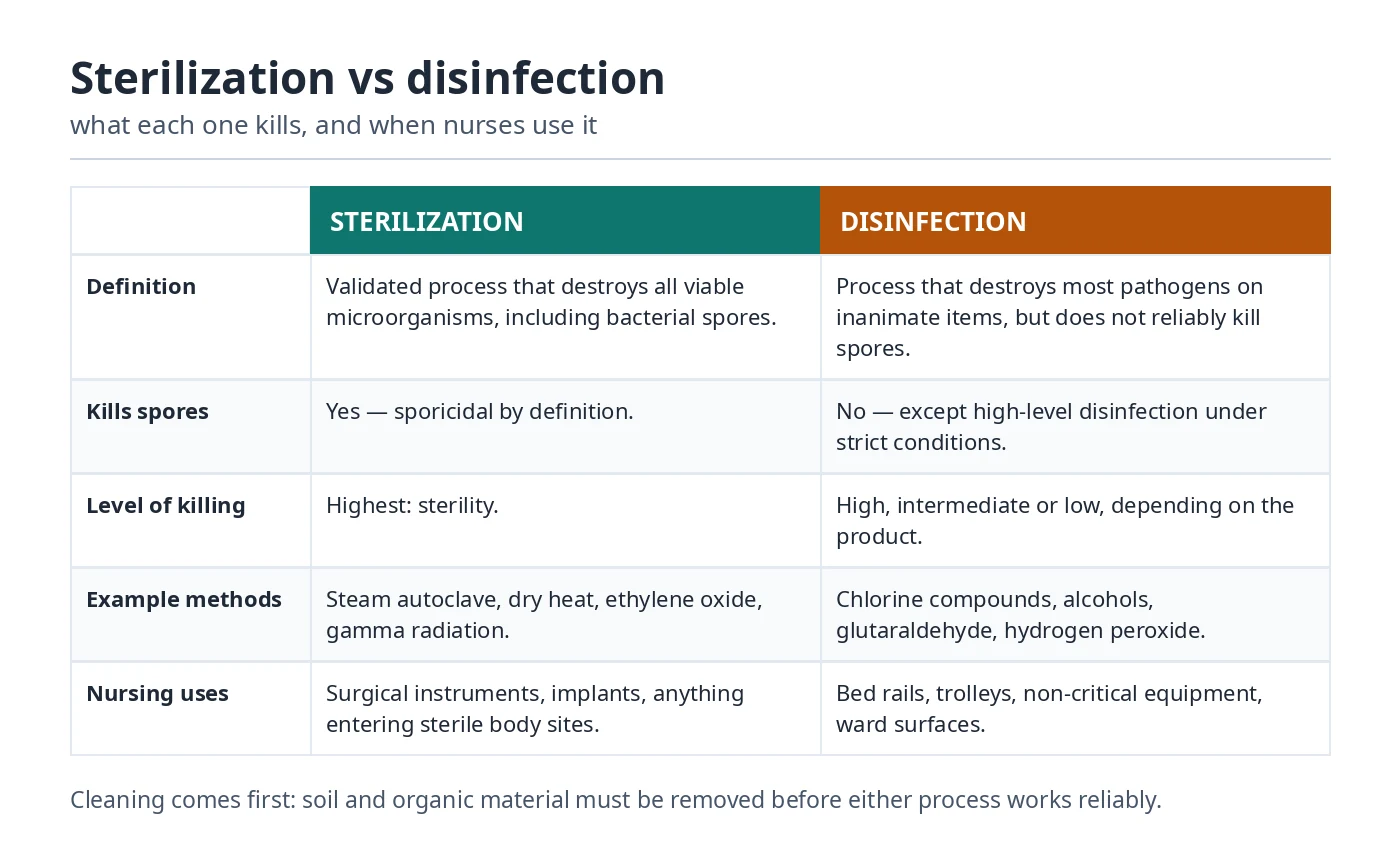
What are the four phases of the bacterial growth curve?
The phases are lag, log (exponential), stationary and death (decline). Bacteria initially adapt, then multiply rapidly, later experience restricted net growth, and finally show a fall in viable cell count. The curve represents a defined batch-culture experiment, so it must not be used as a fixed timeline for disease progression in a patient.
Does a positive bacterial culture always mean antibiotics are needed?
No. A positive culture can indicate infection, colonization or specimen contamination depending on the type of sample and circumstances. A positive urine culture without relevant symptoms is one example in which treatment is often unnecessary, although specific exceptions exist. The clinician makes the treatment decision using patient findings, laboratory evidence and current guidance.
What is the difference between an antigen and an antibody?
An antigen is a molecular target recognized by antibodies or antigen-specific immune receptors. An antibody is an immunoglobulin protein produced by activated B cells or plasma cells and binds a particular target. Antigen tests and antibody tests measure different things; test timing, vaccination history and assay design can all influence what a result means.
How can nurses help reduce antimicrobial resistance?
Nurses help by supporting correct specimen collection, safely administering prescribed medication, documenting and communicating treatment response, recognizing adverse effects and reinforcing appropriate patient education. Infection prevention also reduces the need for antimicrobials. Antibiotics should never be started, withheld or changed independently based solely on a microbiology fact learned from study notes.
Do I need separate chapter pages to study microbiology?
No. The main BSN Microbiology guide provides topic explanations, tables, clinical examples, short and long questions and practice MCQs on one page. Use the topic navigation near the top to jump directly to Gram staining, normal flora, infection prevention or another concept. There is also a dedicated Microbiology MCQ practice page if you prefer questions separately.
Can I use these notes for NCLEX preparation?
They support foundational microbiology knowledge relevant to nursing infection prevention, specimen collection and clinical reasoning. However, the NCLEX also tests broader nursing judgment and current safety standards, so this guide is only one supplementary resource. Use the latest official exam materials and authorized clinical learning resources for comprehensive preparation.
20. Scientific references and further reading
- HEC: Revised BSN/MSN Curriculum (2024), Microbiology course learning outcomes
- OpenStax Microbiology: Staining Microscopic Specimens
- OpenStax Microbiology: How Microbes Grow
- CDC: Core Infection Prevention and Control Practices
- CDC: Transmission-Based Precautions
- CDC NIOSH: Chain of Infection Components
- WHO: Five Moments for Hand Hygiene
Use in clinical placements: These notes explain microbiology principles for study. Actual sample collection, infection prevention, device care and escalation must follow supervised clinical practice and the policies current at your institution.