

Anatomy and Physiology explains how the structures of the human body work together. Anatomy describes body parts, their names and locations. Physiology explains what those parts do and how they respond to changes. This complete BS Nursing learning guide combines Anatomy and Physiology I and II in one searchable article, with two clearly separated semester sections, original explanations, labeled diagrams, examples, answers and practice questions.
Course map: Musculoskeletal, Integumentary, Circulatory and Digestive Systems to Semester 1, and Respiratory, Urinary, Nervous and Sensory, Endocrine and Reproductive Systems to Semester 2. Some universities use a different teaching order, so follow your approved institutional outline when preparing for assessments.
Browse all anatomy topics and semester sections
Find answers to popular Anatomy and Physiology questions
Semester 1 and shared foundations
Semester 2
These questions use the actual searches in the supplied Semrush data. Select a question to jump directly to its explanation. No additional chapter page is needed.
The fundamentals: Anatomical terminology, Cells, Tissues and Homeostasis
What is the difference between Anatomy and Physiology?
Anatomy concerns the structure and location of parts of the body. For example, the heart has four chambers and four valves. Physiology concerns their functions: the chambers receive and pump blood while the valves help maintain directional flow. Nurses need both, because anatomical location helps them assess a patient and physiological knowledge helps them interpret observations.
| Concept | Definition | Example |
|---|---|---|
| Gross anatomy | Structures visible without a microscope | The position of the heart and major bones |
| Microscopic anatomy | Cells and tissues viewed with magnification | Alveolar walls and capillaries |
| Physiology | How an organ or system works | How gas moves across the alveolar membrane |
| Pathophysiology | How disease or injury disrupts function | How reduced cardiac pumping alters tissue perfusion |
Levels of Organization from Cells to Organ Systems
Atoms form molecules, molecules make up cells, similar cells form tissues, several tissues form an organ, and related organs work together in an organ system. For example, cardiac muscle cells form part of heart tissue; the heart is an organ; and the heart and vessels make up the cardiovascular system. A change at a microscopic level can therefore affect the entire body.
Four major tissue classes are epithelial, connective, muscle and nervous tissue. Epithelium lines and protects surfaces. Connective tissue supports, connects and transports material, as seen in bone, cartilage, adipose tissue and blood. Muscle tissue contracts to produce movement. Nervous tissue receives and transmits information.
Anatomical Position, Body Planes and Directional terms
In anatomical position, the person stands upright, faces forward, keeps the arms at the sides and turns the palms forward. Superior means toward the head; inferior means toward the feet; anterior is toward the front; and posterior is toward the back. Medial describes a structure closer to the midline, while lateral is farther away. Proximal is closer to a limb’s point of attachment and distal is farther away.
| Plane | Division | Example of use |
|---|---|---|
| Sagittal | Left and right portions | A sagittal scan shows structures from the side |
| Midsagittal | Equal left and right halves | Describing an organ relative to the midline |
| Frontal or coronal | Front and back portions | A coronal section of the brain |
| Transverse or axial | Upper and lower portions | An axial scan through the abdomen |
Homeostasis, Negative feedback and Positive feedback
Homeostasis is the body’s regulation of internal conditions within ranges compatible with normal function. It does not mean every value stays perfectly constant. Most homeostatic control uses negative feedback: a change is detected, a control mechanism responds and the response opposes the original change. Thermoregulation, blood glucose regulation and many fluid balance processes use this principle.
Worked example: When body temperature rises, receptors and control mechanisms promote heat loss through processes such as skin vasodilation and sweating. As temperature moves toward a regulated range, the drive for cooling decreases. In contrast, positive feedback amplifies a change for a defined purpose, such as the processes surrounding childbirth or blood clot formation.
| Control process | What happens | Examples |
|---|---|---|
| Negative feedback | A response reduces the original deviation | Temperature and blood glucose regulation |
| Positive feedback | A response amplifies the original change until a terminating event | Some stages of labour and clot formation |
| Feedforward control | A response begins in anticipation of a predictable change | Some physiological responses to anticipated activity |
What is homeostasis?
Homeostasis is the regulation of internal conditions within a range that supports normal body function. Body temperature, blood glucose, water balance, blood pH and electrolyte concentrations are regulated through interacting control systems. The controlled values may vary within limits and change predictably with activity, age and clinical condition. Homeostasis does not mean that every body measurement remains constant.
Why is homeostasis important?
Cells depend on a suitable internal environment to carry out metabolism and communicate with one another. Large or persistent changes in temperature, oxygen availability, acidity or electrolyte balance can disrupt enzyme activity, membrane function or organ performance. Nurses study homeostasis so they can understand why physiological observations, symptom trends and laboratory measurements belong to the same clinical picture, even though an abnormal result is not a diagnosis by itself.
What is a negative feedback loop?
Negative feedback is a regulatory process in which a response opposes the change that triggered it. A receptor detects a change, a control center integrates information and an effector produces the response. With an increase in body temperature, sweating and altered blood flow to the skin promote heat loss. Once body temperature moves toward the regulated range, the signals promoting that response diminish. Negative feedback limits deviations rather than reversing every change instantaneously.
Thermoreceptors detect change→
Hypothalamic control response→
Sweating and greater skin heat loss→
Temperature moves toward range
The response reduces the initiating deviation; it is not a positive feedback process.
What is a positive feedback loop?
Positive feedback amplifies an initiating event until another process or a defined endpoint stops it. During childbirth, cervical stretching promotes oxytocin release and uterine contractions; those contractions can cause further stretching. Some parts of clot formation also involve amplification. Positive feedback is less common than negative feedback as a general method of maintaining stable internal conditions.
Semester 1: Anatomy and Physiology I
Follow the BS Nursing sequence: Review the Semester 1 subject directory and Fundamentals of Nursing I alongside these body system explanations.
Semester 1 course focus: Musculoskeletal, integumentary, circulatory and digestive systems. Begin with how the body is built, then examine how it moves, protects itself, transports substances and processes food. This section includes detailed material previously located on the individual femur, radius, vertebrae, tibia and fibula, and lymphatic articles.
1. Skeletal system: Function, Major Bones and Skeletal Divisions
Clinical connection: See body mechanics, range of motion and safe transfers when studying the skeleton’s role in movement.
The adult human skeleton usually contains 206 bones, although small accessory bones and individual anatomical variations can change the count. Bone tissue supports the body, protects organs, assists movement, stores minerals and contains marrow involved in blood cell formation. Bone is living tissue with continuous remodeling by osteoblasts, osteocytes and osteoclasts.
Axial skeleton vs appendicular skeleton
| Axial skeleton | Appendicular skeleton |
|---|---|
| About 80 bones, including the skull, vertebral column and thoracic cage | About 126 bones, including shoulder and pelvic girdles and upper and lower limbs |
| Mainly supports the body’s central framework and protects the brain, spinal cord and thoracic organs | Mainly enables movement, weight transfer and interaction with the environment |
| Examples: cranium, sternum, ribs, vertebrae | Examples: humerus, radius, ulna, femur, tibia, fibula |
Structure of a Long Bone and Bone Tissue
A typical long bone has a shaft called the diaphysis, expanded ends called epiphyses, a compact outer layer, trabecular spongy bone and a medullary cavity. The periosteum covers much of the outer bone surface and the endosteum lines internal surfaces. Articular cartilage covers the joint ends. Growth plates occur in the immature skeleton and normally close with maturation.
Osteoblasts form new bone matrix. Osteoclasts resorb bone, and osteocytes help maintain existing tissue. Calcium and phosphate contribute to the mineral component, while collagen supports resistance to tensile forces. Bone strength reflects both composition and architecture.
2. Joints and Movements: Structure, Classifications and Examples
A joint is a site where bones or skeletal elements meet. Structurally, joints may be fibrous, cartilaginous or synovial. Synovial joints contain a joint cavity, articular cartilage and synovial fluid, and commonly allow a relatively large range of movement. Movement also depends on surrounding muscles, ligaments and other supporting tissues.
| Joint type | Example | Main movement |
|---|---|---|
| Hinge | Elbow, interphalangeal joints | Primarily flexion and extension |
| Ball and socket | Shoulder, hip | Movement in several planes, including rotation |
| Pivot | Proximal radioulnar joint | Rotation |
| Condyloid | Wrist radiocarpal joint | Flexion, extension, abduction and adduction |
| Saddle | Thumb carpometacarpal joint | Movement in two main planes |
| Plane | Some intercarpal joints | Gliding |
Common movement terms include flexion and extension, abduction and adduction, medial and lateral rotation, dorsiflexion and plantar flexion, and pronation and supination. A key exam distinction is that a ligament connects one bone to another, while a tendon connects muscle to bone in the usual teaching example.
3. Muscular System: Muscle types and the Sliding Filament theory
The three main muscle tissue types are skeletal, cardiac and smooth muscle. Skeletal muscle is striated and mainly voluntary, allowing body movement and posture. Cardiac muscle is striated but operates involuntarily within the heart. Smooth muscle lacks obvious striations and is found in the walls of blood vessels, the gastrointestinal tract and other hollow organs.
| Type | Appearance and control | Where it works |
|---|---|---|
| Skeletal | Striated, generally under voluntary control | Attached to the skeleton and other structures |
| Cardiac | Striated, involuntary and electrically connected through specialized junctions | Heart wall |
| Smooth | Non striated, involuntary | Intestines, bladder, blood vessels and uterus |
How does Sliding Filament Theory explain Muscle Contraction?
A skeletal muscle contains bundles of fibers, each made of repeating contractile units called sarcomeres. Within each sarcomere, thin filaments contain actin and thick filaments contain myosin. When a motor neuron activates a muscle fiber, the electrical signal triggers calcium release inside the cell. Calcium binds to troponin, enabling myosin heads to interact with actin. Myosin then uses energy from ATP to cycle through attachment, movement and release, pulling thin filaments toward the sarcomere center. The filaments themselves do not shrink. The sarcomere shortens because their overlap changes.
During contraction, the I band and H zone usually become smaller while the A band remains approximately the same length. ATP is required both for cross bridge cycling and for moving calcium back into storage during relaxation. In clinical learning, this explains why intact nerves, muscle tissue, blood supply, ions and energy availability are all relevant to movement.
How many bones in the human body?
The typical adult human skeleton has 206 bones. About 80 belong to the axial skeleton, which includes the skull, vertebral column and thoracic cage. About 126 belong to the appendicular skeleton, including the limbs and their girdles. Children have more separately identifiable skeletal elements because several bones later fuse. Individual adults can have accessory bones or other anatomical variations, so 206 is a teaching reference, not a count that must fit every person.
What are the three types of muscle tissue?
Skeletal, cardiac and smooth muscle are the three major types. Skeletal muscle produces most voluntary movement and posture. Cardiac muscle contracts rhythmically within the heart, while smooth muscle produces involuntary movement in organs and blood vessel walls. Skeletal and cardiac muscles are striated; smooth muscle is not. The distinction helps explain why different organs contract in different ways and respond to different kinds of regulation.
What is a sarcomere?
A sarcomere is the repeating functional contractile unit within a striated muscle myofibril, bounded by two Z discs. Thin actin filaments extend from the Z discs toward the center, while thick myosin filaments are concentrated in the central region. During contraction, actin slides relative to myosin, decreasing the distance between the Z discs. The A band remains nearly constant, while the I band and H zone become shorter when contraction proceeds.
What is the sliding filament theory?
The sliding filament theory explains how muscle contraction occurs when thin actin filaments move past thick myosin filaments. An electrical signal triggers intracellular calcium release. Calcium binds to troponin, allowing actin and myosin interaction. Myosin cross bridges cycle with the help of ATP, producing force and shortening the sarcomere without shortening the filaments themselves. ATP is also needed for detachment and for the processes that support relaxation.
Muscle fiber excitation→
Calcium release→
Actin and myosin cross bridges→
Sarcomere shortens
| Relaxed sarcomere | Contracting sarcomere |
|---|---|
| Z discs farther apart; I band and H zone wider | Z discs approach; I band and H zone narrow; A band stays approximately the same length |
This is a conceptual study diagram, not a scale drawing of a sarcomere.
How many vertebrae do humans have?
Most adults have 24 movable vertebrae: seven cervical, twelve thoracic and five lumbar. Below these are the sacrum, usually formed from five fused sacral elements, and the coccyx, commonly formed from four fused elements. If the original vertebral elements are counted, the total is commonly around 33. Textbooks use different totals depending on whether they count individual movable bones or fused developmental elements.
4. Femur Bone Anatomy: Labeled Structures and Nursing relevance
Direct answer: The femur is the thigh bone and the longest bone in the human body. Its rounded head articulates with the acetabulum of the pelvis at the hip. Distally, its condyles articulate mainly with the tibia at the knee, and the patellar surface relates to the patella. The femoral neck connects the head to the shaft region.
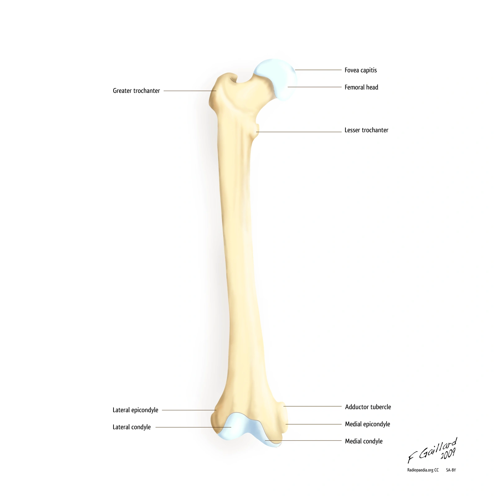

| Region | Landmark | Why it matters |
|---|---|---|
| Proximal femur | Head, neck, greater and lesser trochanters | Hip articulation and muscle attachments |
| Shaft | Diaphysis and linea aspera on posterior surface | Supports load and provides muscle attachment |
| Distal femur | Medial and lateral condyles and epicondyles | Knee articulation and ligament attachments |
In anatomical orientation, the femoral head points medially and the prominent linea aspera lies posteriorly. A condyle is an articular surface, while an epicondyle is a prominence near it where soft tissues attach. Nursing relevance includes recognizing the effects of hip and femoral injuries on mobility, pain, circulation and safe handling.
5. Radius and Ulna: Forearm Landmarks and Pronation
In anatomical position, the radius lies on the thumb side of the forearm, while the ulna lies on the little finger side. The radial head is proximal near the elbow, and the radial styloid process is distal near the wrist. The radius rotates around the ulna during forearm pronation and supination, changing the orientation of the palm.


The radial tuberosity provides attachment for the biceps brachii tendon. Distal radius injuries may affect wrist function and require evaluation of circulation, sensation, movement and pain by qualified clinicians. For exams, remember that the radius is lateral in anatomical position, despite its position changing as the forearm turns.
6. Vertebral Column: Cervical, Thoracic and Lumbar Vertebrae
The typical vertebral column has seven cervical, twelve thoracic and five lumbar movable vertebrae, followed by the sacrum and coccyx. The sacrum usually develops from five fused vertebrae, while the coccyx commonly includes four fused elements, with individual variation. Textbooks may therefore count 24 movable vertebrae, 26 bones including the sacrum and coccyx, or approximately 33 original vertebral elements.


| Region | Typical number | Recognition clue |
|---|---|---|
| Cervical | 7 | Most cervical vertebrae have transverse foramina; C1 atlas and C2 axis are specialized |
| Thoracic | 12 | Articulate with ribs and often have longer downward slanting spinous processes |
| Lumbar | 5 | Large bodies adapted to weight bearing |
| Sacral | Usually 5 fused | Transmits load to the pelvis |
| Coccygeal | Commonly 4 fused | Terminal segment with variable number |
A typical vertebra includes a body, a vertebral arch, spinous and transverse processes, and articular processes. Vertebral foramina collectively help form the canal containing the spinal cord and related structures. Intervertebral foramina are openings through which spinal nerves exit. Spinal curvatures contribute to balance and mechanical load distribution.
7. Tibia and Fibula: Lower Leg Anatomy compared

The tibia is the larger, medial leg bone and carries most weight from the knee toward the ankle. The fibula is smaller and lateral, contributes to ankle stability and provides muscle and ligament attachment. The medial malleolus belongs to the tibia and the lateral malleolus belongs to the fibula.

| Feature | Tibia | Fibula |
|---|---|---|
| Position | Medial, toward body midline | Lateral, toward outer leg |
| Weight bearing | Main weight bearing leg bone | Relatively minor direct weight bearing |
| Knee region | Tibial condyles articulate with femur | Fibular head is near knee but not part of main femorotibial joint |
| Ankle landmark | Medial malleolus | Lateral malleolus |
A trauma assessment may include observation of deformity, swelling, skin integrity, movement, sensation and circulation. Learning the location of each bone helps students describe findings accurately; it does not qualify a student to diagnose fractures from appearance alone.
8. Integumentary System: Layers of Skin, Structures and Functions
Apply skin anatomy to nursing: Explore pressure injury prevention and skin checks, wound assessment and dressing principles, and infection and microorganisms.


What does the Integumentary System do? The skin, hair, nails, sweat glands and sebaceous glands form the integumentary system. It protects against environmental hazards and excess water loss, contributes to temperature regulation, enables sensation and participates in vitamin D synthesis. The skin also plays a role in immune defense and tissue repair.
| Layer | Structure | Main relevance |
|---|---|---|
| Epidermis | Outermost epithelium; includes protective keratinized layers | Barrier protection and limiting water loss |
| Dermis | Connective tissue containing vessels, nerves, follicles and glands | Strength, sensation, nourishment and temperature regulation |
| Hypodermis | Subcutaneous tissue, commonly rich in fat and connective tissue | Insulation, cushioning and tissue attachment |
Epidermis and Dermis: How skin layers differ
The epidermis contains keratinocytes as its most abundant cell type, along with melanocytes, immune related cells and sensory associated cells. It is avascular and receives nutrients by diffusion from underlying tissues. In thick skin, epidermal strata are basale, spinosum, granulosum, lucidum and corneum. The lucidum is especially apparent in palms and soles; thin skin generally lacks a distinct stratum lucidum.
The dermis has papillary and reticular regions that contain collagen and other connective tissue components. Blood vessels, sensory receptors, glands and hair follicles are present here. Sweat evaporation can dissipate heat, while adjustments in skin blood flow also influence heat exchange. Wound healing involves inflammation, tissue formation and remodeling, with the process affected by injury depth, circulation and other factors.
Clinical connection: When assessing skin integrity, distinguish normal variation from a new lesion or pressure related change. Observe site, appearance, symptoms and surrounding tissue rather than assigning an unsupported diagnosis. The detailed dressing procedure belongs in the existing clinical skills library, not this anatomy guide.
What is the integumentary system?
The integumentary system is the skin and its associated structures, including hair, nails, sweat glands and sebaceous glands. It forms the body’s external protective covering and participates in temperature regulation, sensation, immune defense and water balance. The epidermis, dermis and underlying subcutaneous tissue have different structures and clinical significance. The skin is an organ composed of multiple tissue types, not a single layer of cells.
What does the integumentary system do?
The integumentary system helps limit microbial entry and fluid loss, provides mechanical protection, detects touch, pressure, temperature and pain, regulates heat exchange through sweating and changes in skin blood flow, and participates in vitamin D synthesis. Its functions overlap with the cardiovascular, nervous and immune systems. For example, cooling through sweating requires working sweat glands, adequate fluid balance and coordinated neural responses.
How many layers does the skin have?
The skin proper has two major layers: epidermis and dermis. The hypodermis lies underneath and is often shown as a third layer in educational cross sections, but it is subcutaneous tissue rather than part of the skin proper. The epidermis itself has four main strata in most thin skin and five clearly distinguished strata in thick skin. Thick skin is found on the palms and soles and contains a prominent stratum lucidum.
What is the difference between epidermis and dermis?
The epidermis is a keratinized outer epithelial barrier without its own blood vessels. The dermis is a connective tissue layer containing blood vessels, sensory endings, hair follicles and glands. The dermis nourishes the epidermis through diffusion and contributes strength and elasticity. The hypodermis is deeper tissue that often contains adipose cells and helps with insulation, cushioning and attachment to underlying structures.
9. Cardiovascular System: Blood, Heart Anatomy and Circulation
Continue into nursing assessment: Use the heart failure nursing care plan, vital signs and escalation guide, and complete blood count (CBC) explanation to connect circulation with clinical findings.


The cardiovascular system transports oxygen, nutrients, hormones and metabolic waste. Its main components are blood, heart and blood vessels. Arteries generally carry blood away from the heart, veins return it toward the heart, and capillaries allow exchange with tissues. Pulmonary arteries carry blood with relatively low oxygen content, while pulmonary veins usually carry oxygen rich blood.
Heart Anatomy: Four Chambers and Valves
The right atrium receives systemic venous blood through the superior and inferior venae cavae. Blood enters the right ventricle through the tricuspid valve, then passes through the pulmonary valve into the pulmonary trunk. Pulmonary veins return oxygen rich blood to the left atrium. It moves through the mitral valve into the left ventricle, then through the aortic valve into the systemic circulation.
Heart blood flow sequence: Body → venae cavae → right atrium → tricuspid valve → right ventricle → pulmonary valve → pulmonary arteries → lungs → pulmonary veins → left atrium → mitral valve → left ventricle → aortic valve → aorta → body.
The Cardiac Conduction System and Cardiac cycle
The sinoatrial node normally initiates the heartbeat. Electrical activity spreads through the atria toward the atrioventricular node, then through the bundle of His, bundle branches and Purkinje network to coordinate ventricular contraction. Systole describes contraction and blood ejection; diastole describes relaxation and filling. Heart sounds primarily reflect valve closure: S1 is associated with atrioventricular valve closure, and S2 with semilunar valve closure.
Cardiac output formula with worked example
Cardiac output = heart rate × stroke volume. For an example heart rate of 72 beats per minute and a stroke volume of 70 mL per beat, cardiac output is 72 × 70 = 5,040 mL per minute, or approximately 5.0 L/min. This is a calculation example, not a patient treatment target. Cardiac output depends on rate, filling, contractility, vascular resistance and other physiological influences.
| Blood component | Primary function |
|---|---|
| Red blood cells | Transport gases, mainly through hemoglobin |
| White blood cells | Participate in immune defense |
| Platelets | Support primary haemostasis and clot formation |
| Plasma | Liquid medium carrying proteins, electrolytes, nutrients and waste |
The cardiac and vascular system links directly to assessment of pulse, skin perfusion, blood pressure and tissue oxygen delivery. These bedside observations must be interpreted in context rather than as isolated diagnoses.
How does blood flow through the heart?
Blood returns from the body to the right side of the heart, travels to the lungs, returns to the left side of the heart, and is then pumped out to the body. The superior and inferior venae cavae deliver systemic venous blood to the right atrium. Blood passes through the tricuspid valve into the right ventricle, through the pulmonary valve to pulmonary arteries and then to pulmonary capillaries. After gas exchange, pulmonary veins carry blood to the left atrium. It passes the mitral valve to the left ventricle and leaves through the aortic valve and aorta.
Follow the arrows from the body through the lungs and back to systemic circulation.
Blue and red are conceptual oxygenation cues; actual blood is never blue. Diagram not to scale.
What is cardiac output?
Cardiac output is the volume of blood pumped by one ventricle in one minute. It depends on heart rate and stroke volume. Stroke volume is the amount of blood ejected by a ventricle during one beat. The basic formula is cardiac output = heart rate × stroke volume. For a rate of 75 beats per minute and a stroke volume of 70 mL per beat, cardiac output equals 5,250 mL/min, or 5.25 L/min. This is an arithmetic example rather than a target for an individual patient.
How to calculate cardiac output
First, identify the heart rate in beats per minute and the stroke volume in milliliters per beat. Multiply the numbers; the beat unit cancels, leaving milliliters per minute. Finally, divide by 1,000 to convert to liters per minute. For example, 80 beats/min × 65 mL/beat = 5,200 mL/min = 5.2 L/min. If stroke volume is not known, heart rate alone is insufficient to calculate cardiac output.
Which blood vessels carry blood away from the heart?
Arteries carry blood away from the heart, while veins carry blood toward the heart. This directional distinction is more accurate than defining arteries as oxygenated and veins as deoxygenated, because the pulmonary arteries carry relatively oxygen poor blood and the pulmonary veins carry oxygen rich blood. Capillaries have thin walls adapted for exchange of gases, fluids and solutes with tissues.
10. Lymphatic System: Anatomy, Functions and Flow of Lymph
Related learning: The Microbiology nursing guide develops infection and immunity concepts, while CBC blood test notes explain the cells involved in blood and immune responses.
The Lymphatic System is a network of lymphatic capillaries, vessels, nodes, trunks and ducts together with immune associated organs. It returns excess interstitial fluid to venous blood, helps transport absorbed dietary lipids from the intestine, and supports immune surveillance. Lymph moves toward major venous junctions rather than circulating through a heart driven closed loop.


Flow of Lymph: Interstitial fluid → lymphatic capillaries → collecting lymphatic vessels → lymph nodes → lymphatic trunks → lymphatic ducts → venous circulation. The thoracic duct drains most of the body into the left venous angle; the right lymphatic duct typically drains the right upper quadrant into the right venous angle.
A lymph node filters lymph and supports immune recognition. The spleen filters blood and participates in immune responses. The thymus is associated with T lymphocyte maturation. The lymphatic system is relevant to tissue fluid balance and understanding localized swelling, but swelling has multiple possible causes and requires appropriate clinical assessment.
11. Digestive System: Organ Functions, Enzymes and Absorption
Applied topics: Read iron deficiency and anemia for the nutritional consequences of inadequate iron availability, and elderly nursing care challenges for patient-centred considerations around eating and hydration.
The digestive system takes in food, moves it along the gastrointestinal tract, breaks it into smaller components, absorbs nutrients and eliminates undigested material. The alimentary canal runs from mouth through pharynx, oesophagus, stomach, small intestine, large intestine, rectum and anus. Accessory organs include the salivary glands, liver, gallbladder and pancreas.
| Organ or region | Main functions |
|---|---|
| Mouth and salivary glands | Mechanical processing, lubrication and initial starch digestion |
| Oesophagus | Moves swallowed material toward the stomach |
| Stomach | Stores and mixes food; acid and enzymes begin substantial protein digestion |
| Small intestine | Most nutrient digestion and absorption |
| Liver and gallbladder | Bile production and storage/release of bile for fat digestion |
| Pancreas | Delivers digestive enzymes and bicarbonate into the duodenum |
| Large intestine | Water and electrolyte absorption, stool formation and microbiota interactions |
| Rectum and anus | Stool storage and controlled elimination |
What is Peristalsis and how does it move food?
Peristalsis is a coordinated wave of smooth muscle contraction and relaxation that helps move contents along a hollow organ. In the gastrointestinal tract, muscular layers and neural control contribute to propulsion. Segmentation primarily mixes intestinal contents to improve exposure to digestive secretions and absorptive surfaces. The two types of movement work together, but they are not interchangeable.
How does the small intestine absorb nutrients?
The small intestine has circular folds, villi and microvilli that greatly increase available surface area for absorption. Capillaries within villi transport many absorbed nutrients, including most monosaccharides and amino acids, toward the hepatic portal system. Lymphatic lacteals in intestinal villi help transport much of the absorbed long chain fat. The liver receives nutrient rich portal blood and participates in metabolism, storage and detoxification.
Digestion and absorption are related but different: digestion breaks complex food into components that can be absorbed, while absorption transports those components across the intestinal lining into blood or lymph. Nurses connect this knowledge to observing feeding tolerance, hydration, elimination and nutrition related risks.
What is peristalsis?
Peristalsis is a coordinated wave of muscle contraction and relaxation that moves contents through a hollow organ. In the gastrointestinal tract, coordinated circular and longitudinal smooth muscle activity, enteric nerves and other signals move swallowed material and intestinal contents forward. Peristalsis is not the same as segmentation. Segmentation provides repeated mixing of intestinal contents and exposes material to digestive secretions and absorptive surfaces.
How does circulatory system interact with digestive system?
The digestive system breaks down and absorbs nutrients, while the cardiovascular system transports many of those nutrients to the liver and other tissues. Blood capillaries in small intestinal villi receive substances such as absorbed glucose and amino acids. This blood passes through the hepatic portal circulation to the liver, where nutrients are processed, stored or released. The cardiovascular system also delivers oxygen to digestive organs and carries away metabolic waste.
Fat absorption follows an additional route: many long chain dietary lipids are packaged into chylomicrons within intestinal cells and enter lymphatic lacteals. They pass through larger lymphatic vessels before reaching venous circulation. This makes the digestive, lymphatic and cardiovascular systems a particularly useful example of integrated anatomy and physiology.
Small intestinal lumen → intestinal epithelium → villus blood capillaries → hepatic portal vein → liver → systemic blood
Small intestinal lumen → intestinal epithelium → chylomicrons → villus lacteal → lymphatic ducts → venous blood
Simplified physiological pathways. Nutrient transport depends on molecular form and metabolic context.
What does the small intestine do?
The small intestine completes much chemical digestion and performs most nutrient absorption. Its duodenum receives digestive secretions and bile; the jejunum provides a large absorptive surface; and the ileum absorbs selected remaining nutrients, including vitamin B12 bound to intrinsic factor and many bile salts. Circular folds, villi and microvilli increase the surface area available for absorption. The large intestine is particularly important for water and electrolyte reclamation, microbial metabolism and stool formation.
Semester 2: Anatomy and Physiology II
Study pathway: Keep the Semester 2 curriculum directory and Fundamentals of Nursing II available for assessment and clinical skills applications.
Semester 2 course focus: Respiratory, urinary, nervous and sensory, endocrine and reproductive systems. Students should learn how structures support gas exchange, regulate fluids, carry information, control hormones and participate in reproduction. This section integrates the cranial nerve material previously published as a separate revision page.
12. Respiratory System: Organs, Ventilation and Gas Exchange
Related nursing practice: Compare respiratory physiology with lung sounds and auscultation, pneumonia nursing care plans, and respiratory observations and escalation.


The respiratory system moves air into and out of the lungs and allows oxygen to enter blood while carbon dioxide leaves it. Conducting structures include the nose, pharynx, larynx, trachea, bronchi and smaller bronchioles. Gas exchange occurs mainly in the alveoli, which are closely associated with pulmonary capillaries.
Lungs, Pleura and Diaphragm
The right lung generally contains three lobes, while the left has two. The visceral pleura lies on the lung surface and the parietal pleura lines the chest wall. The pleural space contains a thin layer of fluid that permits movement with little friction. The diaphragm is the primary muscle for quiet inspiration, assisted by the external intercostal muscles.
What happens during Inspiration and Expiration?
During quiet inspiration, the diaphragm contracts and thoracic volume increases. Alveolar pressure falls slightly below atmospheric pressure, so air moves inward. During normal quiet expiration, elastic recoil of the lungs and chest wall helps expel air as the inspiratory muscles relax. Forced expiration recruits additional muscles. Air moves according to pressure gradients, not because the lungs actively pull individual oxygen molecules into the bloodstream.
How does gas exchange occur in Alveoli?
Alveoli have thin walls designed for rapid diffusion. Type I pneumocytes provide most of the thin exchange surface; type II pneumocytes produce surfactant, which reduces surface tension and helps maintain alveolar stability. Oxygen diffuses toward pulmonary capillary blood when its partial pressure is higher in alveolar gas, while carbon dioxide usually moves in the opposite direction. Efficient exchange requires adequate ventilation, perfusion and membrane function.
Oxygen is transported primarily by hemoglobin within red blood cells. Carbon dioxide is carried mainly in bicarbonate form, with smaller amounts dissolved or associated with proteins. This distinction explains why breathing rate, ventilation, oxygen saturation and tissue oxygen delivery are related but not identical measurements.
| Lung volume or capacity | Definition |
|---|---|
| Tidal volume | Air moved during a usual quiet breath |
| Inspiratory reserve volume | Additional air inhaled after a normal inspiration |
| Expiratory reserve volume | Additional air exhaled after a normal expiration |
| Residual volume | Air remaining after maximal expiration |
| Vital capacity | Tidal volume + inspiratory reserve + expiratory reserve |
| Total lung capacity | Vital capacity + residual volume |
Normal values vary with body size, age, biological characteristics, measurement technique and clinical status. A usual spirometry test does not directly measure residual volume. In nursing assessment, changes in work of breathing and responsiveness can be clinically important even when an isolated numerical observation appears acceptable.
Which of the following occurs when the diaphragm contracts?
The thoracic cavity expands, intrapulmonary pressure decreases relative to atmospheric pressure, and air flows into the lungs during inspiration. The diaphragm flattens and moves downward as it contracts, increasing vertical thoracic dimension. The external intercostal muscles assist expansion. The pressure difference is small during normal breathing but is sufficient to move air inward. Relaxation and elastic recoil usually account for much of quiet expiration.
How is the cardiovascular system related to the respiratory system?
The respiratory system brings air into contact with the alveoli, where oxygen and carbon dioxide are exchanged with pulmonary capillary blood. The cardiovascular system transports this blood between the lungs and body tissues. The right ventricle pumps blood into pulmonary vessels; the left ventricle pumps oxygenated blood through systemic arteries. Even if the lungs exchange oxygen normally, inadequate circulation can impair its delivery to tissues. Conversely, good cardiac output cannot replace ineffective ventilation or gas exchange.
What is the function of alveoli?
Alveoli are small air spaces whose thin walls are closely associated with pulmonary capillaries. They are the major sites of oxygen and carbon dioxide exchange. Type I alveolar cells contribute a large, thin diffusion surface. Type II cells produce surfactant, reducing surface tension. The diffusion process depends on appropriate partial pressure gradients and the integrity of the air blood barrier. Gas exchange and ventilation are related but different processes.
Oxygen diffuses toward blood
Carbon dioxide arrives from blood
Alveolar epithelium, shared basement membrane regions and capillary endothelium
Oxygen enters red blood cells and binds hemoglobin
Carbon dioxide moves toward the alveolus
Oxygen: alveolus → blood. Carbon dioxide: blood → alveolus. Movement follows partial pressure gradients, not active pumping by the alveoli.
What are lung volumes and capacities?
Lung volumes are individual measurable amounts of air such as tidal volume, inspiratory reserve volume and expiratory reserve volume. Capacities combine two or more volumes. Vital capacity equals tidal volume + inspiratory reserve volume + expiratory reserve volume. Total lung capacity adds residual volume to vital capacity. Students should distinguish the anatomical mechanics of breathing from the physiological measurements used in respiratory testing.
13. Urinary System: Renal Anatomy, Nephron and Urine Formation
Related nursing practice: Learn about urinary catheter care and infection prevention, electrolyte and renal lab values, and hypertonic, hypotonic and isotonic solutions.


The urinary system includes two kidneys, two ureters, the bladder and the urethra. Kidneys help regulate fluid volume, electrolyte composition, acid base status and blood pressure. They also participate in the elimination of metabolic waste and produce hormones involved in red blood cell production and other physiological processes. The ureters carry urine to the bladder, where it is stored until voiding.
Renal Cortex, Medulla and Urine collection
The kidney’s outer cortex surrounds the medulla, which contains renal pyramids. Collecting ducts drain urine toward the papillae, minor and major calyces, renal pelvis and ureter. The nephron is the functional unit of the kidney. It includes the renal corpuscle, proximal tubule, loop of Henle, distal tubule and connecting and collecting structures. The renal corpuscle contains glomerular capillaries and Bowman’s capsule.
| Nephron component | Main function |
|---|---|
| Glomerulus and Bowman’s capsule | Filter water and small solutes from plasma while normally retaining blood cells and most proteins |
| Proximal tubule | Reabsorb much of the filtered water, sodium, glucose, amino acids and bicarbonate |
| Loop of Henle | Help establish the concentration gradient needed for urine concentration |
| Distal tubule | Adjust the handling of selected electrolytes |
| Collecting duct | Support hormone regulated water and electrolyte handling |
Filtration, Reabsorption and Secretion explained
Filtration moves water and small dissolved substances from glomerular blood into the nephron. Reabsorption transfers needed substances and water from tubular fluid back toward blood. Secretion moves selected substances from blood into tubular fluid. Excretion represents the remaining amount eliminated through urine. A useful conceptual relationship is: excretion = filtration − reabsorption + secretion.
Renin Angiotensin Aldosterone System and ADH
The kidneys release renin in response to appropriate signals involving renal perfusion, sodium delivery and sympathetic activity. Renin initiates a pathway forming angiotensin I and then angiotensin II. Angiotensin II contributes to vasoconstriction and stimulates aldosterone release. Aldosterone promotes sodium reabsorption and potassium secretion in selected distal nephron segments.
Antidiuretic hormone (ADH), produced in the hypothalamus and released from the posterior pituitary, helps regulate water conservation by altering collecting duct water permeability. ADH mainly regulates water reabsorption, while aldosterone mainly changes sodium and potassium transport. Both influence the overall balance of extracellular fluid.
Nursing application: Understanding these pathways helps students interpret why fluid balance, urine output, blood pressure and electrolyte monitoring can be related. Abnormal observations require appropriate clinical assessment rather than being diagnosed from a textbook chart.
What is a nephron?
A nephron is the microscopic functional unit of the kidney that filters blood plasma and modifies the resulting fluid to form urine. Each nephron includes a renal corpuscle with a glomerulus and Bowman’s capsule, together with a system of renal tubules. Millions of nephrons across the kidneys collectively help regulate water, electrolyte concentration, acid base balance and waste elimination. The collecting duct system works with nephrons to determine the final concentration of urine.
What are the three steps of urine formation?
The three main processes are glomerular filtration, tubular reabsorption and tubular secretion. Filtration transfers water and many small solutes from glomerular blood into Bowman’s space. Reabsorption moves useful substances back from tubular fluid to blood. Secretion adds selected substances from blood into tubular fluid. The amount excreted in urine reflects the balance of these processes and the regulation of water handling.
Filtration to Bowman’s space →
Proximal tubule
Major water and solute reabsorption →
Loop of Henle
Concentration gradient →
Distal tubule
Selective ion regulation →
Collecting duct
ADH and aldosterone effects →
Urine
This is a simplified flow sequence, not a drawing of the nephron’s true three dimensional shape. Reabsorption and secretion occur in multiple segments.
What is RAAS?
RAAS means the renin angiotensin aldosterone system. It is a hormonal pathway that contributes to blood pressure, sodium balance and extracellular fluid regulation. Renin released by the kidney helps produce angiotensin I from angiotensinogen. Angiotensin converting enzyme contributes to the formation of angiotensin II. Angiotensin II can affect blood vessels and stimulate aldosterone release from the adrenal cortex. Aldosterone promotes sodium conservation and potassium secretion in the distal nephron.
What is the difference between ADH and aldosterone?
ADH mainly increases water reabsorption by raising water permeability in the collecting ducts, while aldosterone primarily increases sodium reabsorption and potassium secretion in selected distal nephron segments. Water can follow retained sodium under appropriate physiological conditions. Both help regulate fluid balance, but ADH is not simply another name for aldosterone, and their triggers and receptor pathways differ.
14. Nervous System: Neurons, the Brain, Reflex Arc and Autonomic Control
Clinical reasoning: Extend nervous system anatomy with the stroke types and nursing observations guide and Next Generation NCLEX clinical judgment case study.
The central nervous system consists of the brain and spinal cord. The peripheral nervous system includes nerves and ganglia outside these structures. Sensory pathways carry information toward the central nervous system, while motor pathways carry commands to effectors. The autonomic nervous system controls many involuntary functions and includes sympathetic, parasympathetic and enteric components.
Structure of a Neuron and Function of Myelin
Neurons contain a cell body, dendrites and an axon. Dendrites receive signals, and the axon transmits activity toward other cells. Many axons are wrapped in myelin, which helps impulses travel efficiently through saltatory conduction between nodes of Ranvier. Supporting glial cells help maintain the neural environment, insulation and defense.
Action potential and Synaptic transmission
In a typical neuron, reaching a threshold can open voltage gated sodium channels, allowing sodium to enter and depolarize the membrane. Sodium channel inactivation and potassium movement contribute to repolarization. The sodium potassium pump maintains long term ion gradients but does not produce every phase of the rapid electrical signal. At many chemical synapses, an arriving impulse triggers calcium dependent release of neurotransmitter, which binds receptors on the next cell.
Reflex Arc: Five Parts and their Roles
A reflex is a relatively rapid, patterned response to a stimulus. In a common five part teaching model, a receptor detects the stimulus, a sensory neuron carries the signal, an integration center processes it, a motor neuron carries a response signal and an effector produces the response. Some simple reflexes can be organized at spinal level while still being influenced by higher neural pathways.
| Reflex component | Function |
|---|---|
| Receptor | Detects the stimulus |
| Sensory neuron | Carries information toward the central nervous system |
| Integration center | Processes signals in spinal cord or brain |
| Motor neuron | Carries output toward the target |
| Effector | Produces a response such as muscle contraction |
Brain Lobes, Cerebellum and Brainstem
The frontal lobe supports executive functions and voluntary movement. Parietal regions process body sensations and spatial information. The temporal lobe contributes to hearing, memory and language functions, and the occipital lobe plays a central role in vision. The cerebellum supports coordination, balance and motor learning. The brainstem includes pathways and nuclei important to breathing, arousal and other essential functions.
The meninges are dura mater, arachnoid mater and pia mater. Cerebrospinal fluid circulates through ventricular and subarachnoid compartments, providing mechanical protection and other support.
Sympathetic vs Parasympathetic Nervous System
| Sympathetic tendency | Parasympathetic tendency |
|---|---|
| Often prepares the body for increased activity and stress | Often supports rest, recovery and routine maintenance |
| Commonly raises heart rate through appropriate receptors | Vagal influence commonly slows sinoatrial node activity |
| May reduce gastrointestinal movement during acute stress | Often supports gastrointestinal activity |
| Effects vary by organ and receptor subtype | Effects vary by organ and receptor subtype |
What is the somatic nervous system?
The somatic nervous system is the part of the peripheral nervous system concerned with sensory information from the body and voluntary control of skeletal muscles. Somatic motor neurons innervate skeletal muscle fibers, allowing movements such as walking or reaching. Somatic sensory pathways carry information from skin, muscles and joints toward the central nervous system. Many reflexes also use somatic pathways, so somatic does not mean that every response is consciously initiated.
What is a reflex arc?
A reflex arc is the neural pathway involved in a relatively rapid, patterned response to a stimulus. A basic five component model includes a receptor, sensory neuron, integration center, motor neuron and effector. A knee jerk reflex illustrates a relatively simple spinal circuit involving muscle stretch receptors, sensory fibers and motor output. Withdrawal responses can involve additional interneurons and more complex circuitry.
Detects stimulus → 2. Sensory neuron
Carries signal inward → 3. Integration center
Processes signal → 4. Motor neuron
Carries output → 5. Effector
Produces response
Simplified neural pathway for learning. Individual reflexes vary in their number of synapses and neural connections.
What is an action potential?
An action potential is a rapidly propagating electrical change in the membrane potential of an excitable cell. In a typical neuron, voltage gated sodium channels allow inward sodium movement during the rising phase, followed by sodium channel inactivation and increased potassium conductance during repolarization. An action potential is all or none at the local membrane region once threshold conditions are met. Stronger stimulation is generally encoded by patterns and frequencies of neural activity, not by making each action potential much taller.
What is the difference between sympathetic and parasympathetic nervous systems?
The sympathetic and parasympathetic divisions are components of autonomic regulation. Sympathetic pathways often coordinate responses to activity and stress, such as increasing heart rate and changing vascular tone. Parasympathetic pathways often support digestion, gland secretion and slowing of the heart through vagal activity. Both divisions operate continually, and their effects depend on the organ and receptor involved. It is misleading to describe one system as always active and the other as always switched off.
15. The Twelve Cranial Nerves: Names, Functions and Memory Guide
There are twelve pairs of cranial nerves, numbered I through XII. Their functions include smell, vision, eye movements, facial sensation, facial movement, hearing, balance, swallowing, speech, shoulder movement and tongue control. Learning them supports anatomical understanding of clinical assessment, but abnormal findings need interpretation by qualified clinicians.
| Nerve | Name | Main function | Type |
|---|---|---|---|
| I | Olfactory | Smell | Sensory |
| II | Optic | Vision | Sensory |
| III | Oculomotor | Most eye movements, eyelid elevation and pupil constriction | Motor and parasympathetic |
| IV | Trochlear | Superior oblique muscle | Motor |
| V | Trigeminal | Facial sensation and chewing muscles | Mixed |
| VI | Abducens | Lateral rectus muscle | Motor |
| VII | Facial | Facial expression, anterior tongue taste and some gland functions | Mixed |
| VIII | Vestibulocochlear | Hearing and balance | Sensory |
| IX | Glossopharyngeal | Posterior tongue taste and swallowing functions | Mixed |
| X | Vagus | Voice, swallowing and wide parasympathetic functions | Mixed |
| XI | Accessory | Trapezius and sternocleidomastoid movement | Motor |
| XII | Hypoglossal | Tongue movement | Motor |
Remember the groups by function: I and II are special senses; III, IV and VI involve eye movements; V and VII relate strongly to face sensation and expression; VIII concerns hearing and balance; IX and X contribute to swallowing and voice; XI controls selected neck and shoulder muscles; XII controls tongue movement.
Cranial Nerves Mnemonic and Sensory, Motor, mixed grouping
To remember the twelve cranial nerve names in order, use the first letters O O O T T A F V G V A H. The original revision phrase from our earlier notes is: “Our Observant Owl Travels Through Alpine Forests, Visiting Gardens, Valleys And Hills.” Repeat the letters while pointing to the nerve numbers I through XII. The phrase is only a memory cue and does not describe nerve function.
In the traditional introductory grouping, cranial nerves I, II and VIII are sensory; III, IV, VI, XI and XII are mainly motor; and V, VII, IX and X are mixed. Actual cranial nerve fiber functions can be more nuanced than these broad labels.

16. Sensory Systems: Eye Anatomy, Hearing and Balance
Anatomy of the Human Eye and how Vision works
Light passes through the transparent cornea, aqueous humor, pupil, lens and vitreous body to reach the retina. Retinal photoreceptors transduce light into signals that are processed through retinal neurons and then travel along the optic nerve. Rods are especially sensitive in dim lighting, while cones contribute to colour discrimination and high acuity. The fovea has high cone density and supports detailed central vision.
The iris regulates the amount of light entering the eye by changing pupil size. Ciliary muscle activity alters lens shape during accommodation for near or distant focus. The pupil light reflex involves optic sensory input and oculomotor pathways. Changes in pupils should be described accurately and assessed according to clinical protocols.
Ear Anatomy: Sound transmission and Vestibular function
The external ear collects sound and directs it toward the tympanic membrane. The middle ear ossicles are the malleus, incus and stapes. Their movement transmits vibrations toward the inner ear. In the cochlea, hair cells convert mechanical vibration into neural signals that travel via the cochlear part of cranial nerve VIII.
The vestibular system includes semicircular canals and otolith organs, which sense aspects of head rotation and linear movement. Vision, proprioception and vestibular input work together to maintain balance. Hearing and balance share cranial nerve VIII but use distinct sensory receptors.
17. Endocrine System: Glands, Hormones and Regulation
Related study resources: Connect endocrine regulation to the Type 2 diabetes nursing care plan and interpreting lab results for nurses.
Endocrine glands release hormones that travel through body fluids, commonly blood, and act on cells bearing appropriate receptors. The hypothalamus, pituitary, thyroid, parathyroids, adrenal glands, pancreatic islets, pineal gland and gonads are major contributors to endocrine regulation. Hormones may act on metabolism, growth, reproduction, water balance, stress responses and other processes.
| Gland | Selected hormones | Principal physiology |
|---|---|---|
| Anterior pituitary | GH, TSH, ACTH, FSH, LH and prolactin | Growth, regulation of other glands and reproductive functions |
| Posterior pituitary | Releases ADH and oxytocin made in the hypothalamus | Water conservation and some reproductive functions |
| Thyroid | T3 and T4 | Metabolic regulation and development |
| Parathyroids | Parathyroid hormone | Calcium regulation |
| Adrenal cortex | Cortisol, aldosterone and adrenal androgens | Metabolism, stress response and electrolyte regulation |
| Adrenal medulla | Epinephrine and norepinephrine | Rapid sympathetic related effects |
| Pancreatic islets | Insulin and glucagon | Blood glucose control |
| Gonads | Estrogens, progesterone and testosterone | Reproductive development and function |
Insulin vs Glucagon
Insulin is secreted mainly by pancreatic beta cells and generally promotes nutrient uptake and storage while reducing circulating glucose. Glucagon is secreted mainly by pancreatic alpha cells and promotes hepatic glucose output under appropriate conditions. Their overall effects help maintain blood glucose availability. Glucose balance also depends on food intake, activity and several additional hormonal mechanisms.
Endocrine Negative feedback example
The hypothalamus releases thyrotropin releasing hormone, which stimulates TSH release from the anterior pituitary. TSH stimulates thyroid hormone synthesis and release. Thyroid hormones generally feed back on the hypothalamus and pituitary, reducing further stimulation when levels are sufficiently high. This is a useful example of how a feedback loop regulates an organ system.
What does the pituitary gland do?
The pituitary gland releases hormones that influence growth, thyroid activity, adrenal function, reproduction, lactation and water balance. Its anterior and posterior parts have different physiological roles. The anterior pituitary synthesizes hormones including GH, TSH, ACTH, FSH, LH and prolactin. The posterior pituitary releases ADH and oxytocin, which are synthesized in the hypothalamus. Although called the master gland in introductory teaching, the pituitary is itself regulated by hypothalamic signals and feedback from target organs.
Where is the pituitary gland located?
The pituitary is a small endocrine gland at the base of the brain, seated within the sella turcica of the sphenoid bone and connected to the hypothalamus by the infundibulum. The pituitary lies close to the optic chiasm, an anatomical relationship that helps explain why particular disorders affecting nearby structures may be associated with changes in vision. The location is an anatomical fact, not a guide to diagnosing a patient.
What is the difference between insulin and glucagon?
Insulin is produced primarily by pancreatic beta cells and supports nutrient uptake and storage, generally lowering circulating glucose. Glucagon is produced primarily by alpha cells and helps raise circulating glucose through effects on hepatic metabolism, particularly during fasting. These hormones interact with stress hormones, diet, tissue insulin sensitivity and other mechanisms. They form an important example of homeostatic regulation rather than two permanently opposed on/off switches.
TRH → Anterior pituitary
TSH → Thyroid gland
T3 and T4 → Target tissues
As circulating thyroid hormone levels rise, negative feedback generally decreases further stimulation at the hypothalamus and pituitary.
18. Reproductive System: Anatomy, Gametogenesis and Hormonal Cycles
Continue learning: Review the maternal and newborn nursing foundation guide to connect reproductive anatomy with nursing education.
The male reproductive system includes testes, epididymides, ductus deferens, accessory glands, penis and scrotum. The female reproductive system includes ovaries, uterine tubes, uterus, cervix, vagina and external genital structures, collectively called the vulva. Both systems have reproductive and endocrine functions. Sensitive topics should be taught respectfully without assuming a patient’s gender, beliefs or reproductive goals.
Spermatogenesis: Stages of Sperm Formation
Sperm develop in seminiferous tubules of the testes. Spermatogonia give rise to cells that undergo meiosis and maturation, ultimately producing haploid sperm cells. Sertoli cells provide support to developing cells, while Leydig cells produce testosterone. Sperm undergo additional functional maturation in the epididymis. The process requires appropriate hormonal regulation and suitable tissue conditions.
Oogenesis: Stages and Comparison with Spermatogenesis
Oogenesis begins during fetal development. Primary oocytes enter meiosis but pause for an extended period. During reproductive years, selected follicles progress through development during ovarian cycles. A secondary oocyte commonly pauses in metaphase II, with completion of meiosis II occurring after fertilization. Unlike sperm production, oogenesis typically produces one large functional gamete rather than four similarly sized mature cells from a single meiosis.
| Feature | Spermatogenesis | Oogenesis |
|---|---|---|
| Usual onset | Puberty | Before birth |
| Location | Testes | Ovaries |
| Output per completed meiosis | Usually four haploid spermatids | Usually one functional ovum and polar bodies |
| Pattern | Ongoing production in typical adult physiology | Cyclical maturation through reproductive years |
| Completion | Meiosis is completed during sperm development | Meiosis II usually finishes after fertilization |
Ovarian and Menstrual cycles explained
The ovarian cycle includes follicular development, ovulation and a luteal phase. Follicle stimulating hormone supports follicular development, and a midcycle rise in luteinizing hormone ordinarily triggers ovulation. The corpus luteum secretes progesterone after ovulation, helping support the uterine secretory phase. If implantation does not occur, luteal hormone levels generally decline and menstruation follows.
The uterine cycle is often described as menstrual, proliferative and secretory phases. A 28 day cycle is a teaching example rather than a fixed normal length, because cycle timing varies across individuals and life stages. Fertilization most commonly occurs in the ampullary part of a uterine tube, and implantation begins after early embryonic development.
What is oogenesis?
Oogenesis is the developmental process that produces female gametes within the ovaries. It begins before birth when oogonia give rise to primary oocytes that enter meiosis and pause in prophase I. Following puberty, selected follicles develop as part of ovarian cycles. Usually one dominant follicle progresses toward ovulation in a cycle. The secondary oocyte is released after completion of meiosis I and typically remains arrested in metaphase II until fertilization triggers completion of meiosis II.
What is spermatogenesis?
Spermatogenesis is the formation of sperm cells in the seminiferous tubules of the testes. Diploid spermatogonia give rise to primary spermatocytes that enter meiosis, then to haploid spermatids that mature into spermatozoa. Sertoli cells support developing cells while Leydig cells produce testosterone. Unlike the developmental pattern of oogenesis, sperm production begins at puberty and usually continues throughout much of adult life, though rate and quality can vary.
Diploid (2n) → Primary oocyte
Arrest in prophase I → Secondary oocyte
Haploid (n), arrest in metaphase II → Fertilization
Completion of meiosis II
Polar bodies are also formed during unequal meiotic divisions. This is a teaching sequence, not a literal cellular image.
What are the phases of the menstrual cycle?
The menstrual cycle can be described from the perspective of ovarian activity and uterine endometrial changes. Ovarian phases include follicular development, ovulation and the luteal phase. Endometrial phases are often described as menstrual, proliferative and secretory. Rising estradiol helps build the endometrium during the proliferative phase, while progesterone from the corpus luteum supports secretory changes. The commonly taught 28 day cycle is illustrative; a person’s actual cycle length and ovulation timing can vary considerably.
19. Nursing Applications: Connecting Anatomy to Patient Observations
From anatomy to nursing care: Study Fundamentals of Nursing I and II, nursing care plans and worked examples and the clinical procedures and skills library.
| System | What anatomy and physiology explain | Nursing learning connection |
|---|---|---|
| Bones and muscles | Support, movement and physical alignment | Describe injury location, assist with approved movement and understand basic mobility |
| Skin | Protective barrier, receptors and temperature responses | Recognize and communicate changes in skin integrity |
| Heart and blood | Pumping, blood flow and tissue delivery | Understand pulse, perfusion and relevant circulation observations |
| Digestive | Motility, digestion and absorption | Understand nutrition, feeding tolerance and elimination |
| Respiratory | Ventilation, diffusion and gas transport | Recognize changes in breathing effort and oxygenation |
| Urinary | Filtration, reabsorption, secretion and fluid regulation | Understand fluid balance charts and urine formation |
| Nervous and sensory | Signaling, reflexes and sensory perception | Describe changes in movement, alertness and sensation |
| Endocrine | Hormonal signaling and feedback | Understand physiological reasons for some laboratory and symptom patterns |
| Reproductive | Organ structure, gamete development and cycles | Support accurate, respectful health education |
Clinical safety: These are educational explanations of normal human structure and function. Clinical assessment, diagnosis, medicines and procedures must follow the relevant professional scope, qualified supervision and current institutional guidance.
Clinical Reasoning and Visual Practice: Connect Body Systems
These are educational examples of anatomy and physiology, not instructions for independent clinical diagnosis. Read the scenario, explain the underlying structures and then review the answer.
Case 1. Why can a Hip Injury Reduce Walking Ability?
Situation: An older adult reports difficulty bearing weight after a fall. Learning explanation: The hip transfers load between the pelvis and femur, while surrounding ligaments and muscles contribute to stability and movement. Pain and an injured weight bearing structure can limit function. Recognizing femoral head, neck and trochanter landmarks helps describe the anatomical location. A patient with suspected injury should not be encouraged to walk without appropriate clinical assessment.
Case 2. Why does loss of skin Integrity affect more than appearance?
Situation: A patient has a large damaged skin area. Learning explanation: The epidermis normally limits water loss and entry of external organisms, while dermal vessels and receptors support perfusion, sensation and temperature regulation. Damage can therefore affect fluid conservation, protection and comfort. The nursing focus is accurate assessment and appropriate escalation, not assigning a diagnosis from an illustration.
Case 3. How are the Heart and Lungs connected?
Situation: A student observes a change in breathing effort and pulse. Learning explanation: Airflow brings oxygen to alveoli, diffusion transfers oxygen to pulmonary blood, and the left heart sends that oxygen carrying blood through systemic vessels. Ventilation, diffusion, circulation and hemoglobin are all necessary for effective oxygen delivery. No single observation establishes whether all these processes are functioning normally.
Case 4. Why might a change in kidney function affect fluids and electrolytes?
Situation: Fluid measurements and laboratory results change during a hospital stay. Learning explanation: Nephrons filter plasma, return useful materials to blood and secrete selected substances. Hormones such as ADH and aldosterone modify water and electrolyte handling. Understanding these pathways helps a nurse recognize why fluid records and laboratory values are related. Interpretation must account for the entire clinical picture.
Case 5. How do Nerves and Muscles work together when moving a limb?
Situation: A person attempts to bend the elbow. Learning explanation: Motor pathways send signals to skeletal muscle fibers. Calcium mediated interaction of actin and myosin produces tension, which transmits through tendons to bones and joints. Sensory input and the central nervous system help coordinate the resulting movement. Impairment in muscles, nerves, joints or blood supply may affect function.
Blank diagram challenge for exam revision
After studying the labeled figures, cover the labels and try these tasks on paper: identify eight major landmarks of the femur, label the radius and ulna in anatomical position, locate the five regions of the vertebral column, draw the route of blood through all four heart chambers, outline a nephron from Bowman’s capsule to collecting duct, and name all twelve cranial nerves in order. Explain the function of each structure rather than memorizing its position alone.
Revision check: A useful answer should state the correct anatomical structure, its function, a relevant physiological mechanism and one implication for patient assessment. If one element is missing, revisit the section above before moving to questions.
20. Short answer questions for Anatomy and Physiology I and II
Semester 1: Ten short questions with answers
What is the difference between Anatomy and Physiology?
Anatomy studies the body’s structure and position. Physiology studies how those structures work. Heart chambers are anatomy; pumping and cardiac output are physiology.
How many bones are in a typical Adult Skeleton?
Usually 206, although accessory bones and individual variation can change the count. The axial skeleton has about 80 bones and the appendicular skeleton about 126.
Which is lateral in Anatomical Position, Radius or Ulna?
The radius is lateral on the thumb side; the ulna is medial on the little finger side.
Name the main regions of the Vertebral column.
Seven cervical, twelve thoracic and five lumbar vertebrae form the 24 commonly counted movable vertebrae, followed by the sacrum and coccyx.
What is the difference between Tibia and Fibula?
The tibia is medial and bears most leg weight. The fibula is lateral and helps with muscle attachment and ankle stability.
What is the difference between the Epidermis and Dermis?
The epidermis is an avascular outer epithelial barrier. The dermis is vascular connective tissue containing many nerves, glands and follicles.
What does Cardiac output mean?
The volume of blood pumped by one ventricle per minute. Cardiac output equals heart rate multiplied by stroke volume.
What is the difference between Pulmonary and Systemic Circulation?
Pulmonary circulation carries blood between the right heart and lungs and returns it to the left heart. Systemic circulation carries blood from the left heart to body tissues and returns it to the right.
What are the main functions of the Lymphatic system?
Returning interstitial fluid to blood, transporting many absorbed dietary fats and supporting immune surveillance.
How does Peristalsis differ from Segmentation?
Peristalsis mainly propels contents through the digestive tract. Segmentation mixes intestinal contents and helps digestion and absorption.
Semester 2: Ten short questions with answers
Where does oxygen enter blood in the lungs?
Across the alveolar capillary membrane, driven by partial pressure differences between alveolar gas and capillary blood.
What is the primary muscle of quiet inspiration?
The diaphragm. Its contraction enlarges the thoracic cavity and creates a pressure gradient for air entry.
Name the three processes of urine formation.
Glomerular filtration, tubular reabsorption and tubular secretion.
What is the difference between ADH and aldosterone?
ADH supports water conservation by increasing collecting duct water permeability. Aldosterone mainly affects sodium reabsorption and potassium secretion.
What are the components of a reflex arc?
A receptor, sensory neuron, integration center, motor neuron and effector.
Which cranial nerve provides vision?
Cranial nerve II, the optic nerve. Vision also requires healthy retinal receptors, neural pathways and visual cortical processing.
Which cranial nerve carries hearing and balance information?
Cranial nerve VIII, the vestibulocochlear nerve.
How does a sympathetic response differ from parasympathetic activity?
Sympathetic pathways commonly support increased activity and stress responses, while parasympathetic pathways often support rest, digestion and recovery. Effects vary by organ.
Which gland releases TSH?
The anterior pituitary. TSH stimulates thyroid hormone production, while thyroid hormones participate in feedback regulation.
How does oogenesis differ from spermatogenesis?
Oogenesis begins before birth and commonly results in one functional ovum per completed meiosis; spermatogenesis begins at puberty and generally produces four haploid spermatids per meiosis.
21. Anatomy and Physiology MCQs with explained answers
More practice: Use the nursing MCQ library and NCLEX practice questions with rationales after completing this article’s questions.
Choose an answer before opening the explanation. These are original nursing study questions, not official PNMC past examination papers. They are separated by semester and use ordinary accessible HTML instead of visual cards.
Semester 1: Original anatomy MCQs
1. Which structure forms the outer epithelial layer of the skin?
- Dermis
- Epidermis
- Hypodermis
- Periosteum
Show answer and explanation
Correct: B — Epidermis. The epidermis is the outer skin layer and contributes to its protective barrier.
2. A tendon primarily connects which structures?
- Bone to bone
- Muscle to bone
- Nerve to skin
- Artery to vein
Show answer and explanation
Correct: B — Muscle to bone. Tendons transmit forces from muscle to bone, enabling movement.
3. What is the main mechanical role of the heart?
- Produce bile
- Pump blood through pulmonary and systemic circuits
- Absorb dietary nutrients
- Filter urine
Show answer and explanation
Correct: B — Pump blood through pulmonary and systemic circuits. The heart generates the flow needed for blood circulation.
4. When body temperature rises, sweating promotes cooling. This is an example of:
- Positive feedback
- Negative feedback
- Irreversible adaptation
- Genetic mutation
Show answer and explanation
Correct: B — Negative feedback. Sweating opposes the original rise in temperature, a negative-feedback response.
5. Where is most nutrient absorption from digested food carried out?
- Oesophagus
- Stomach
- Small intestine
- Large intestine
Show answer and explanation
Correct: C — Small intestine. The small intestine is specialised for nutrient digestion and absorption.
6. Which type of vessel generally carries blood away from the heart?
- Vein
- Lymphatic duct
- Artery
- Capillary
Show answer and explanation
Correct: C — Artery. Arteries carry blood away from the heart; oxygen content depends on the circuit.
7. A patient has extensive loss of the skin barrier. Which physiological function is most directly threatened?
- Mechanical protection and limitation of fluid loss
- Red-blood-cell synthesis in bone marrow
- Insulin release from the pancreas
- Bile formation in the liver
Show answer and explanation
Correct: A — Mechanical protection and limitation of fluid loss. The skin barrier protects tissues and reduces excessive water loss; barrier damage also raises infection risk.
8. A negative-feedback loop fails to restore a variable toward its expected range. Which explanation most directly describes the problem?
- The effector did not adequately oppose the deviation
- A receptor never participates in feedback
- All feedback mechanisms amplify change
- The control centre has no role in physiology
Show answer and explanation
Correct: A — The effector did not adequately oppose the deviation. Negative feedback requires sensing, integration and an effective counter-response.
9. Nutrients absorbed from the small intestine enter blood and are delivered to the liver through which pathway?
- The hepatic portal circulation
- The pulmonary vein directly
- The ureter
- The coronary sinus
Show answer and explanation
Correct: A — The hepatic portal circulation. The hepatic portal system carries nutrient-rich blood from many digestive organs to the liver before systemic return.
10. What is the primary role of peristalsis?
- Produce bile
- Move contents through the digestive tract
- Filter blood in the kidneys
- Store glucose in the liver
Show answer and explanation
Correct answer: B. Coordinated smooth muscle contractions propel material through the digestive system.
11. Which vessels return oxygen rich blood from the lungs to the left atrium?
- Pulmonary arteries
- Pulmonary veins
- Venae cavae
- Aorta
Show answer and explanation
Correct answer: B. Pulmonary veins transport blood from the lungs to the left atrium. Vessel names reflect the direction of flow relative to the heart, not oxygen content.
Additional Semester 1 question 1. Which part of a sarcomere remains nearly the same length during contraction?
- A band
- I band
- H zone
- Distance between Z discs
Show answer and explanation
Correct answer: A. Thick filament length remains approximately constant, so the A band does not shorten like the I band and H zone.
Additional Semester 1 question 2. Which structure normally contains the stratum lucidum?
- Thick skin of palms and soles
- Thin scalp skin
- Dermis
- Hypodermis
Show answer and explanation
Correct answer: A. A distinct stratum lucidum is a characteristic of thick skin, particularly the palms and soles.
Additional Semester 1 question 3. Which tissue directly forms the contractile walls of the heart?
- Cardiac muscle
- Smooth muscle
- Skeletal muscle
- Adipose tissue
Show answer and explanation
Correct answer: A. The myocardium contains cardiac muscle cells that generate the force for pumping.
Additional Semester 1 question 4. Which valve lies between the left atrium and left ventricle?
- Mitral valve
- Tricuspid valve
- Pulmonary valve
- Aortic valve
Show answer and explanation
Correct answer: A. The mitral valve, also called the bicuspid valve, separates the left atrium and ventricle.
Additional Semester 1 question 5. Cardiac output is calculated as:
- Heart rate × stroke volume
- Blood pressure × body mass
- Pulse × temperature
- Vital capacity × respiratory rate
Show answer and explanation
Correct answer: A. The formula combines the volume ejected per beat with the number of beats per minute.
Additional Semester 1 question 6. Which region primarily absorbs most digested nutrients?
- Small intestine
- Oesophagus
- Stomach
- Rectum
Show answer and explanation
Correct answer: A. Villi and microvilli give the small intestine a very large absorptive surface.
Additional Semester 1 question 7. Which fluid pathway returns much absorbed long chain fat?
- Intestinal lacteals and lymphatic vessels
- Renal tubules
- Pulmonary arteries
- Cardiac conduction pathways
Show answer and explanation
Correct answer: A. Many absorbed long chain lipids enter lymphatic lacteals as chylomicrons.
Additional Semester 1 question 8. What is the main purpose of peristalsis?
- Propel intestinal contents
- Produce insulin
- Filter blood
- Conduct sound
Show answer and explanation
Correct answer: A. Peristaltic waves help move material along the digestive tract.
Additional Semester 1 question 9. Which is lateral in anatomical position?
- Radius relative to ulna
- Tibia relative to fibula
- Sternum relative to ribs
- Sacrum relative to ilium
Show answer and explanation
Correct answer: A. The radius lies on the thumb side of the forearm, while the ulna is medial.
Additional Semester 1 question 10. Which part of a typical vertebra forms the large anterior weight bearing area?
- Vertebral body
- Spinous process
- Transverse foramen
- Intervertebral foramen
Show answer and explanation
Correct answer: A. The vertebral body bears much of the load, particularly in the lumbar region.
Additional Semester 1 question 11. Which option correctly names the three major muscle tissue types?
- Skeletal, cardiac and smooth
- Axial, appendicular and synovial
- Epithelial, osseous and neural
- Red, white and yellow
Show answer and explanation
Correct answer: A. Muscle tissue is classified as skeletal, cardiac or smooth based on its structure and function.
Additional Semester 1 question 12. Which response is a negative feedback example?
- Sweating in response to a rise in body temperature
- Amplifying contractions during labour
- Continued reinforcement of clotting
- Uncontrolled increasing temperature without response
Show answer and explanation
Correct answer: A. Sweating can promote heat loss, opposing the rise in body temperature.
Semester 2: Application and recall MCQs
1. Where does most gas exchange occur in the lungs?
- Alveoli
- Trachea
- Pharynx
- Pleural cavity
Show answer and explanation
Correct answer: A. Alveoli have thin membranes adjacent to capillaries that facilitate diffusion.
2. What helps reduce surface tension within alveoli?
- Surfactant
- Bile
- Renin
- Insulin
Show answer and explanation
Correct answer: A. Surfactant from type II alveolar cells helps stabilize the alveoli.
3. Which nephron structure performs initial blood filtration?
- Glomerulus
- Ureter
- Bladder
- Renal pelvis
Show answer and explanation
Correct answer: A. Filtration occurs across glomerular capillaries into Bowman’s space.
4. Which process returns filtered glucose to blood under normal conditions?
- Tubular reabsorption
- Secretion
- Micturition
- Glomerular filtration alone
Show answer and explanation
Correct answer: A. Most filtered glucose is ordinarily reabsorbed in the proximal tubule.
5. Which hormone directly regulates collecting duct water permeability?
- ADH
- Oxytocin
- TSH
- Glucagon
Show answer and explanation
Correct answer: A. ADH helps regulate water conservation in the collecting duct.
6. Which structure is responsible for much of conscious visual processing?
- Occipital cortex
- Cerebellar vermis
- Medulla oblongata
- Pituitary gland
Show answer and explanation
Correct answer: A. The occipital cortex plays a central role in cortical vision.
7. Which cranial nerve supplies the lateral rectus muscle?
- Abducens nerve VI
- Optic nerve II
- Vagus nerve X
- Accessory nerve XI
Show answer and explanation
Correct answer: A. Cranial nerve VI innervates the lateral rectus muscle.
8. Which structure participates in the hearing receptor system?
- Cochlea
- Semicircular canal only
- Thyroid
- Retina
Show answer and explanation
Correct answer: A. Cochlear hair cells transduce sound vibrations into neural signals.
9. Which pituitary hormone stimulates thyroid hormone secretion?
- TSH
- ADH
- Oxytocin
- Prolactin
Show answer and explanation
Correct answer: A. Thyroid stimulating hormone acts on the thyroid gland.
10. Which hormone is secreted mainly by pancreatic beta cells?
- Insulin
- Glucagon
- Aldosterone
- Epinephrine
Show answer and explanation
Correct answer: A. Insulin is produced by pancreatic beta cells.
11. Where does fertilization commonly take place?
- Ampulla of uterine tube
- Cervical canal
- Ovary
- Vaginal canal
Show answer and explanation
Correct answer: A. Fertilization most often occurs in the ampullary part of a uterine tube.
12. In a typical reflex arc, what detects the stimulus?
- Receptor
- Motor neuron
- Effector
- Adrenal gland
Show answer and explanation
Correct answer: A. A receptor responds to the relevant stimulus.
13. What is a normal function of the cerebellum?
- Movement coordination
- Secretion of insulin
- Bile formation
- Urine storage
Show answer and explanation
Correct answer: A. The cerebellum contributes to balance, movement coordination and motor learning.
14. Which part of the urinary tract stores urine?
- Bladder
- Kidney cortex
- Ureter
- Urethra
Show answer and explanation
Correct answer: A. The urinary bladder temporarily stores urine before micturition.
15. Which process usually triggers ovulation?
- LH surge
- Decline of all hormones
- ADH secretion
- Renin release
Show answer and explanation
Correct answer: A. A midcycle surge in luteinizing hormone normally initiates ovulation.
Additional Semester 2 question 1. What happens when the diaphragm contracts during quiet inspiration?
- Thoracic volume increases
- Thoracic volume falls sharply
- Alveoli stop exchanging gases
- Air is pushed out immediately
Show answer and explanation
Correct answer: A. Diaphragm contraction increases thoracic volume and helps produce the pressure gradient for air entering the lungs.
Additional Semester 2 question 2. Which cell produces pulmonary surfactant?
- Type II alveolar cell
- Red blood cell
- Goblet cell of the trachea
- Myelinated neuron
Show answer and explanation
Correct answer: A. Type II pneumocytes produce surfactant, which reduces alveolar surface tension.
Additional Semester 2 question 3. In the kidney, most filtered glucose is usually reabsorbed in the:
- Proximal tubule
- Bladder
- Ureter
- Renal pelvis
Show answer and explanation
Correct answer: A. Glucose is ordinarily reabsorbed in the proximal tubule by carrier-mediated transport.
Additional Semester 2 question 4. Which hormone increases water permeability in the collecting duct?
- Antidiuretic hormone
- Thyroid stimulating hormone
- Insulin
- Prolactin
Show answer and explanation
Correct answer: A. ADH regulates aquaporin insertion and water reabsorption in the collecting duct.
Additional Semester 2 question 5. Which action describes tubular secretion?
- Movement from peritubular blood into tubular fluid
- Movement from tubules back to blood
- Movement of urine from bladder to urethra
- Movement of blood through the renal artery
Show answer and explanation
Correct answer: A. Secretion transfers selected substances from blood into the tubule.
Additional Semester 2 question 6. Which reflex arc component carries information toward the spinal cord?
- Sensory neuron
- Motor neuron
- Effector muscle
- Adrenal cortex
Show answer and explanation
Correct answer: A. Afferent sensory neurons convey receptor information toward central neural processing.
Additional Semester 2 question 7. Which part of a neuron carries action potentials toward the terminal?
- Axon
- Dendrite
- Nucleus
- Golgi apparatus
Show answer and explanation
Correct answer: A. Action potentials normally propagate along the axon toward presynaptic terminals.
Additional Semester 2 question 8. Which cranial nerve carries hearing and balance information?
- Vestibulocochlear VIII
- Hypoglossal XII
- Accessory XI
- Olfactory I
Show answer and explanation
Correct answer: A. Cranial nerve VIII comprises sensory pathways for hearing and balance.
Additional Semester 2 question 9. Which gland releases TSH?
- Anterior pituitary
- Posterior pituitary
- Thyroid gland
- Adrenal medulla
Show answer and explanation
Correct answer: A. TSH is synthesized and secreted by the anterior pituitary.
Additional Semester 2 question 10. What hormone is typically released by pancreatic beta cells?
- Insulin
- Glucagon
- Aldosterone
- Cortisol
Show answer and explanation
Correct answer: A. Beta cells secrete insulin, which contributes to regulation of nutrient availability.
Additional Semester 2 question 11. In oogenesis, a secondary oocyte usually completes meiosis II after:
- Fertilization
- Menstruation begins
- The follicle first forms
- Menopause
Show answer and explanation
Correct answer: A. The secondary oocyte ordinarily finishes meiosis II after sperm entry.
Additional Semester 2 question 12. Which event normally triggers ovulation?
- Luteinizing hormone surge
- Erythropoietin release
- Renin secretion
- Sodium reabsorption
Show answer and explanation
Correct answer: A. The midcycle LH surge ordinarily triggers ovulation.
22. Important Anatomy and Physiology long answer questions
Each model answer demonstrates the concepts expected in a structured response. Use the detailed sections and labeled diagrams above to explain structures clearly in your own words.
Semester 1: Six long answer models
1. Explain anatomical position, body planes and homeostasis.
Model answer: In anatomical position, the body is upright, facing forward, with arms alongside the trunk and palms forward. Superior and inferior describe relative position along the head to foot axis; medial and lateral refer to the midline, while proximal and distal describe position along a limb. Sagittal, frontal and transverse planes divide the body in three orientations. Homeostasis describes regulation of internal conditions. Negative feedback opposes a deviation through coordinated receptor, control and effector responses. For example, a rise in body temperature stimulates responses that can increase heat loss. These concepts help nurses document locations and understand changes in physiology.
2. Discuss the axial and appendicular skeleton, long bone structure, and major landmarks.
Model answer: The adult skeleton is usually described as 206 bones, with about 80 axial and 126 appendicular bones. The axial skeleton includes skull, vertebral column and thoracic cage. Appendicular structures include limb bones and girdles. A long bone has a shaft, epiphyses, compact and trabecular bone, articular cartilage and marrow. Bone remodels through osteoblast, osteocyte and osteoclast activity. Explain that the femur has a head and neck, trochanters and distal condyles; the radius lies on the thumb side of the forearm; and the tibia lies medial to the fibula. Relate each landmark to joint function and accurate anatomical description.
3. Explain sliding filament theory and compare the main muscle tissues.
Model answer: Skeletal, cardiac and smooth muscle differ in their structure and control. Skeletal and cardiac tissues are striated, whereas smooth muscle is not. Skeletal muscle fibers contain sarcomeres with actin and myosin filaments. A motor nerve impulse initiates calcium release, exposing binding sites and enabling cross bridge cycling. ATP supports myosin function and relaxation. During contraction, overlapping filaments slide relative to one another so that the sarcomere shortens. The filaments do not shrink. Cardiac muscle supports pumping, and smooth muscle supports movements in hollow organs and vessels. Nursing relevance includes the effects of impaired movement on mobility, function and safety.
4. Describe the integumentary system and its role in barrier protection.
Model answer: The integumentary system comprises skin and associated hair, nails and glands. The avascular epidermis forms an outer epithelial barrier; the dermis contains connective tissue, vessels, sensory structures and many glands; subcutaneous tissue provides cushioning and insulation. Keratin contributes to barrier function, sweat evaporation supports cooling, and cutaneous vessels influence heat exchange. Skin participates in sensation and immune defense. The epidermis regenerates through keratinocyte activity, while deeper injury involves a complex healing response. Explain how nurses use structural knowledge to describe skin changes objectively and recognize when further assessment is required.
5. Trace blood through the heart and explain the cardiac cycle and cardiac output.
Model answer: Deoxygenated systemic venous blood returns to the right atrium through the venae cavae and passes the tricuspid valve into the right ventricle. It flows through the pulmonary valve and arteries to the lungs. Oxygen rich blood returns via pulmonary veins to the left atrium, passes the mitral valve into the left ventricle, and leaves through the aortic valve to systemic circulation. The sinoatrial node normally initiates electrical activity that reaches the AV node and ventricular conduction system. Systole describes ventricular contraction and ejection, while diastole describes relaxation and filling. Cardiac output is heart rate multiplied by stroke volume. For example, 72 beats/min × 70 mL/beat = 5.04 L/min.
6. Describe how digestion, absorption and lymphatic transport work together.
Model answer: Digestive organs include the mouth, oesophagus, stomach, intestines, liver, gallbladder and pancreas. Mechanical and chemical processes break food into absorbable components. Peristalsis moves contents along the digestive tract, while segmentation contributes to mixing. The small intestine has folds, villi and microvilli to increase its absorptive surface. Most absorbed monosaccharides and amino acids enter blood and travel toward the liver through portal circulation. Much absorbed long chain fat enters intestinal lymphatic lacteals and travels through lymphatic vessels toward the venous circulation. This links gastrointestinal physiology with the lymphatic system’s role in fluid return and fat transport.
Semester 2: Four long answer models
1. Explain ventilation, diffusion and gas transport in the respiratory system.
Model answer: Air passes through conducting airways toward the alveoli. During inspiration, the diaphragm contracts and thoracic volume increases, helping establish the pressure gradient for airflow. Quiet expiration largely reflects elastic recoil. At alveoli, oxygen and carbon dioxide diffuse across a thin membrane according to partial pressure differences. Type I alveolar cells provide much of the exchange surface; type II cells produce surfactant. Oxygen travels mainly bound to hemoglobin, while most carbon dioxide is transported as bicarbonate. Explain that effective oxygen delivery depends on ventilation, lung perfusion, gas exchange and blood circulation. Respiratory observations require clinical context and trends.
2. Explain nephron anatomy, urine formation and hormone effects.
Model answer: The nephron includes the renal corpuscle, proximal tubule, loop of Henle, distal tubule and associated collecting structures. Glomerular filtration transfers water and small solutes from blood to tubular fluid. Reabsorption returns needed substances to circulation; secretion transfers selected compounds into tubular fluid. The loop of Henle helps create the medullary concentration gradient. ADH increases collecting duct water permeability, while aldosterone primarily changes sodium and potassium transport in distal nephron regions. The renin angiotensin aldosterone system responds to signals related to perfusion and salt handling. Relate these processes to fluid balance and why intake/output alone cannot establish a diagnosis.
3. Describe neuronal signaling, a reflex arc and important cranial nerves.
Model answer: A neuron has dendrites, a cell body and an axon. Ion channel activity generates action potentials, and synapses transmit information to other cells. The central nervous system comprises brain and spinal cord; the peripheral nervous system connects it with body structures. A five component reflex arc includes a receptor, sensory neuron, integration center, motor neuron and effector. Identify key cranial nerves: II for vision, III/IV/VI for major eye movements, V for facial sensation and chewing, VII for facial expression, VIII for hearing and balance, IX/X for swallowing and speech related functions, XI for selected neck and shoulder movement, and XII for tongue movement. Clinical assessment remains a supervised activity.
4. Describe major endocrine glands, hormonal feedback and reproductive physiology.
Model answer: Endocrine glands produce hormones that act on cells with corresponding receptors. The hypothalamus and pituitary coordinate many hormonal axes, including the thyroid and reproductive systems. Insulin and glucagon contribute to glucose regulation. Negative feedback limits excessive stimulation, as seen when thyroid hormones reduce hypothalamic and pituitary signals. In reproduction, meiosis produces haploid gametes. Spermatogenesis begins at puberty and can proceed continuously, while oogenesis starts before birth and features prolonged meiotic arrest. Ovarian hormones coordinate follicular development, ovulation and luteal events with menstrual changes in the uterine lining. Explain these topics respectfully and distinguish normal physiology from diagnosis of symptoms.
23. Frequently asked Anatomy and Physiology questions
Are Anatomy and Physiology 1 and 2 separate courses?
Yes. They are distinct semester courses with different sets of body systems, but the notes are consolidated on this page for easy navigation.
Which body systems are in Anatomy and Physiology I?
The 2024 HEC and PNMC BSN curriculum focuses on musculoskeletal, integumentary, circulatory and digestive systems, alongside homeostasis and nursing applications.
Which body systems are in Anatomy and Physiology II?
It covers the respiratory, urinary, nervous and sensory, endocrine and reproductive systems.
How can I memorize bones without confusing their locations?
Start with anatomical position, learn axial versus appendicular divisions, then compare landmarks and relations. Label a blank skeleton and practise orientation with proximal, distal, medial and lateral terms.
What is the difference between radius and ulna?
In anatomical position, the radius lies laterally on the thumb side and the ulna medially on the little finger side. The radius moves around the ulna during pronation and supination.
What is the difference between tibia and fibula?
The tibia is the larger medial leg bone and bears most weight; the fibula is lateral and supports muscle attachments and ankle stability.
Why do textbooks give different vertebrae counts?
They may count the 24 individual movable vertebrae, 26 adult bones including fused sacrum and coccyx, or about 33 developmental vertebral elements. Coccygeal numbers vary.
What is the main function of the integumentary system?
It provides a protective barrier, limits water loss, supports sensation, temperature regulation, immune defense and vitamin D synthesis.
How does blood flow through the heart?
Blood returns from the body to the right heart, travels to the lungs, returns to the left heart and is pumped back to systemic tissues. The four cardiac valves promote directional flow.
How do I calculate cardiac output?
Multiply heart rate by stroke volume. For example, 72 beats/min multiplied by 70 mL/beat gives approximately 5.0 L/min.
What is peristalsis?
A coordinated wave of smooth muscle activity that helps propel contents through the gastrointestinal tract.
What is the difference between alveoli and bronchioles?
Bronchioles conduct air within the lungs, while alveoli are the small air spaces closely involved in gas exchange.
How many steps are involved in urine formation?
The three broad processes are glomerular filtration, tubular reabsorption and tubular secretion. The final amount excreted depends on all three.
What is a reflex arc?
A pathway from stimulus detection through neural processing to an effector response, commonly taught as receptor, sensory neuron, integration center, motor neuron and effector.
What are the 12 cranial nerves?
They are the olfactory, optic, oculomotor, trochlear, trigeminal, abducens, facial, vestibulocochlear, glossopharyngeal, vagus, accessory and hypoglossal nerves.
Which gland controls other endocrine glands?
The pituitary releases hormones regulating several endocrine glands, although it is itself regulated by the hypothalamus and feedback from target glands.
What is oogenesis?
The developmental formation of female gametes, beginning before birth and progressing through meiosis with long periods of arrest.
Are these notes official PNMC exam questions?
No. They are educational study notes and original practice questions mapped to broad curriculum concepts, not an official examination question bank.
24. Curriculum, scientific references and image credits
Hero illustration: Ryan Hoyme (massagenerds), Skeleton and muscles, made available under CC0.

Body system and semester navigation icons: Twemoji graphics, CC BY 4.0. The illustrations aid navigation and do not replace the labeled anatomy diagrams in the learning material.
For curriculum alignment, consult the HEC and PNMC revised BSN curriculum. For scientific explanations, consult OpenStax Anatomy and Physiology 2e, NCBI Bookshelf and institutionally recommended anatomy and physiology textbooks. The femur, radius, vertebral column and lymphatic images are attributed in their captions with licensing and source information.
Study navigation: Return to Semester 1 notes · Return to Semester 2 notes · Short answers · MCQs · Semester 1 directory · Semester 2 directory. The information required to revise all systems is provided on this page.